
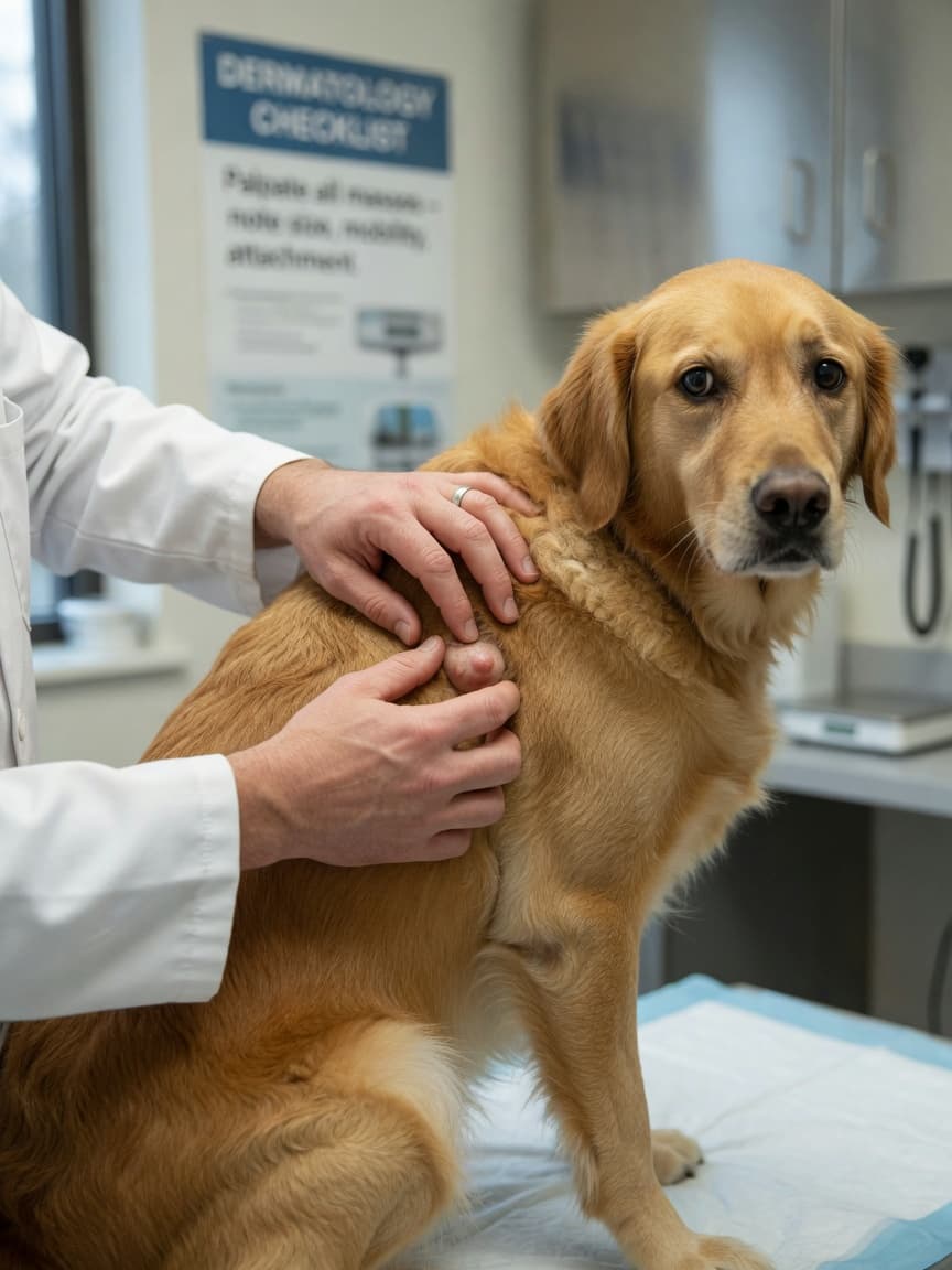
Should You Be Worried About a Lump on Your Dog?
Dogs seem to develop lumps and bumps with remarkable regularity, and for many owners, discovering one is an anxious moment. The good news is that a significant proportion of skin growths in dogs are benign — but the important caveat is that you cannot tell this by looking or feeling alone. Some of the most dangerous tumours can appear deceptively normal, while some alarming-looking lumps turn out to be entirely harmless. The only way to know for certain is veterinary assessment, often with a simple diagnostic test. Here is an overview of the most common types, what distinguishes them, and when to act urgently.
The Most Common Types of Lumps and Bumps in Dogs
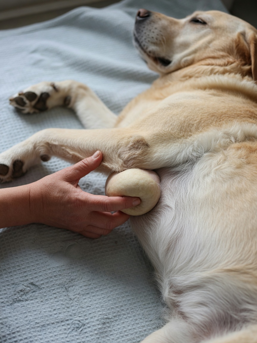
Lipomas (Fatty Lumps)
Lipomas are the most frequently encountered lumps in dogs and are benign tumours of fat cells. They typically feel soft, smooth, and moveable under the skin, like a small bag of soft dough, and are usually found on the chest, abdomen, and upper legs. They grow slowly, do not cause pain when touched, and have well-defined edges. Lipomas are particularly common in middle-aged to older dogs and in overweight individuals. Certain breeds — including Labrador Retrievers, Weimaraners, and Cocker Spaniels — appear to be predisposed.
Most lipomas require no treatment unless they grow in a location that impairs movement, such as in the armpit or groin, or become very large. However, because a rare malignant variant called a liposarcoma exists — and because a firm, infiltrative lipoma can be difficult to distinguish from more serious growths — any new fatty-feeling lump should still be assessed by your vet.
Sebaceous Cysts
Sebaceous cysts form when a skin gland becomes blocked, resulting in a smooth, round, dome-shaped lump just beneath the skin surface. They are usually small — between a few millimetres and a centimetre — and may appear white or slightly bluish through the skin. Occasionally they rupture, releasing a thick, white or grey, cheese-like material. While this looks alarming, it is generally not dangerous, though ruptured cysts can become infected and may require veterinary attention. Intact cysts are usually left alone unless they are causing problems, as surgical removal is straightforward if needed.
Warts (Papillomas)
Canine papillomas are caused by the canine papillomavirus and appear as rough, cauliflower-like growths, often found around the mouth, eyes, or between the toes. They are most common in young dogs and immunosuppressed dogs. In young, healthy dogs, papillomas typically resolve on their own within a few months as the immune system mounts a response. They are not usually harmful, though large or numerous warts in or around the mouth can occasionally interfere with eating, and secondary infection is possible.
Histiocytomas
Histiocytomas are benign tumours of immune cells that appear rapidly — sometimes seemingly overnight — as small, pink, button-like raised lumps, typically on the head, ears, or limbs. They are particularly common in young dogs under two years of age and in certain breeds including Flat-Coated Retrievers and Boxers. Despite their alarming sudden appearance, histiocytomas almost always regress spontaneously within one to three months without any treatment. That said, because their appearance can be similar to mast cell tumours (see below), veterinary confirmation is strongly advisable.
Mast Cell Tumours — The Great Mimics
Mast cell tumours (MCTs) are one of the most common and most serious skin tumours in dogs, accounting for around 20 percent of all canine skin tumours. They are sometimes called "the great mimics" because they can look and feel like almost anything — a soft fatty lump, a firm nodule, a raised pink growth, or an ulcerated sore. MCTs release histamine and other chemicals, and some dogs show surrounding redness or swelling, or signs of gastric irritation such as vomiting and dark stools.
Certain breeds are at significantly higher risk, including Boxers, Labrador Retrievers, Golden Retrievers, Staffordshire Bull Terriers, and Pugs. Mast cell tumours are graded — Grade I tumours confined to the skin have an excellent prognosis with surgical removal, while higher-grade tumours that have spread to lymph nodes or internal organs carry a much more guarded outlook. Early diagnosis is therefore crucial. Never assume a lump is benign simply because it seems soft or small.
Melanomas
Melanomas in dogs are tumours arising from pigment-producing cells. Cutaneous melanomas — those on haired skin — are often benign, especially if small and pigmented. However, melanomas found in the mouth, on the toes, or at mucocutaneous junctions (where skin meets mucous membrane) are frequently malignant and can spread rapidly to lymph nodes and the lungs. Any darkly pigmented or ulcerating growth in these locations warrants urgent veterinary assessment.
Enlarged Lymph Nodes
Lymph nodes are not technically skin lumps, but enlarged nodes — found under the jaw, in front of the shoulders, in the armpits, in the groin, and behind the knees — are a common reason owners bring their dog to the vet concerned about a "lump." Lymph nodes can swell in response to local infection, immune activation, or — most seriously — lymphoma, one of the most common cancers in dogs. Symmetrically enlarged, firm, painless lymph nodes across multiple sites are a red flag for lymphoma and require prompt investigation.
Understanding Diagnostic Tests
The first-line diagnostic tool for most lumps is a fine needle aspirate (FNA). This is a quick, usually painless procedure — no sedation required in most cases — in which your vet inserts a small needle into the lump to collect a sample of cells. These cells are then examined under a microscope (cytology). FNA is very useful for diagnosing lipomas, mast cell tumours, cysts, and lymphoma, though it cannot always provide a definitive answer.
For a more detailed diagnosis — particularly when the FNA is inconclusive or when surgical planning is needed — a biopsy may be recommended. This involves removing a core or wedge of tissue under sedation or anaesthesia and sending it to a specialist pathologist for histopathology. Histopathology provides information not just on the cell type but on the tumour grade, margins, and likely behaviour.
The Rules of Thumb: When to Act Urgently
Any lump should be brought to your vet's attention, but the following signs indicate the lump needs urgent assessment rather than watchful waiting:
- Rapid growth — a lump that doubles in size within a few weeks
- Changes in colour, particularly darkening or the appearance of new pigment
- Ulceration or bleeding from the lump surface
- A hard, irregular, or fixed (non-moveable) lump
- A lump on or near the mouth, toes, or a mucocutaneous junction
- Signs of systemic illness — vomiting, weight loss, lethargy, or swollen lymph nodes elsewhere in the body
- Any lump in a high-risk breed, particularly Boxers, Bulldogs, or Retrievers
When watchful waiting may be appropriate — for example, a small, soft, slow-growing lump in an otherwise healthy young dog — always establish a clear review plan with your vet and document the lump with photographs and measurements so that any changes are easy to detect. The BSAVA advises that any lump growing consistently, even slowly, should be investigated rather than simply monitored indefinitely.