
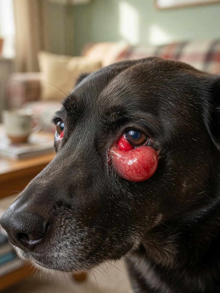
What Is Cherry Eye?
If you've noticed a red, fleshy lump appearing in the inner corner of your dog's eye, you're likely looking at cherry eye. The formal name is prolapse of the nictitans gland, and whilst it looks alarming, it is a condition that responds well to treatment when addressed promptly.
The third eyelid — the nictitating membrane — is a thin, protective structure that sweeps across the eye from the inner corner. Nestled at its base is a tear gland responsible for producing a significant proportion of your dog's tear film. In dogs prone to cherry eye, the connective tissue anchoring this gland is weak, allowing the gland to pop out of position and become visible as that characteristic red mass.
Which Dogs Are Most at Risk?
Cherry eye has a clear breed predisposition. It most commonly affects brachycephalic breeds — those with flattened facial anatomy — and certain other breeds with loose facial tissue. Dogs frequently diagnosed with cherry eye include Bulldogs, Beagles, Cocker Spaniels, Shih Tzus, Lhasa Apsos, Basset Hounds, and Boston Terriers.
The condition is particularly common in young dogs, typically under two years of age, as the connective tissue is still developing. However, it can occur at any age. Both eyes can be affected, either simultaneously or one after the other, so owners who have had one eye treated should keep a close watch on the second.
What Causes Cherry Eye?
The underlying cause is a weakness or laxity in the retinaculum — the fibrous tissue that holds the third eyelid gland in place. This is believed to be largely genetic in predisposed breeds, which is why the condition tends to run in family lines. Once the gland prolapses, exposure to air and the environment causes it to become irritated, swollen, and sometimes infected, which makes it even more resistant to returning to its correct position on its own.
Trauma, rubbing, or vigorous play can trigger the initial prolapse, but in susceptible dogs, the gland was already primed to shift position. There is no reliable way to prevent cherry eye in a dog with the underlying genetic predisposition.
Symptoms to Watch For
The primary sign is the obvious red or pink oval mass visible in the inner corner of the eye. Beyond the visual, you may notice:
- Increased tear production or watery discharge
- Rubbing or pawing at the affected eye
- Mild conjunctival redness around the mass
- Mucous discharge if secondary infection develops
- Squinting or apparent discomfort
The gland itself is not painful in the early stages, but prolonged exposure leads to swelling, dryness, and discomfort. In chronic cases, the gland can sustain permanent damage that impairs its ability to produce tears even after surgical correction.
Why Prompt Treatment Matters
The third eyelid gland produces approximately 30 to 50 per cent of a dog's total tear film. This makes it far too important to simply remove, which was once standard practice before vets understood the long-term consequences. Dogs that had the gland removed as a routine treatment for cherry eye had significantly elevated rates of dry eye (keratoconjunctivitis sicca) later in life — a chronic condition requiring daily management and carrying a risk of vision impairment.
Modern treatment focuses on preserving the gland and returning it to its correct anatomical position. The sooner this is done after prolapse, the better the prognosis for long-term gland function.
Treatment Options
Surgical Repositioning
Surgery is the standard and most effective treatment for cherry eye. The goal is to return the prolapsed gland to its pocket and secure it so it cannot prolapse again. There are two main surgical techniques used by veterinary ophthalmologists and experienced general practitioners.
The pocket technique (also called the tucking technique) involves creating a small pocket of tissue over the gland and suturing it closed, effectively burying the gland back in position. This is currently considered the gold standard approach and carries a high success rate.
The anchoring technique involves suturing the gland directly to the periorbita or orbital rim to hold it in position. It is used in some cases where the pocket technique is not feasible, though recurrence rates can be slightly higher depending on technique and individual anatomy.
Can Cherry Eye Resolve Without Surgery?
In rare cases, particularly in very young puppies, gentle massage of the prolapsed gland may allow it to slip back into position temporarily. Some vets may attempt this alongside anti-inflammatory medication as a short-term measure. However, the underlying ligamentous laxity remains, and the gland almost invariably prolapses again. Surgery remains the only reliable long-term solution.
Recovery After Cherry Eye Surgery
Recovery is generally smooth and uncomplicated. Dogs go home the same day in most cases. Expect a period of one to two weeks of restricted activity, with instructions to prevent your dog from rubbing the eye — an Elizabethan collar is essential during this time.
Post-operative care typically includes antibiotic eye drops or ointment to prevent infection and anti-inflammatory medication to reduce swelling. Swelling around the eye is normal in the first few days and should gradually subside.
Follow-up appointments allow the vet to check that sutures are holding and the gland is settling correctly. Success rates with the pocket technique exceed 90 per cent in skilled hands, though a small proportion of dogs require a second procedure if the gland re-prolapses.
Long-Term Outlook
Most dogs that undergo timely cherry eye surgery go on to have normal tear production and comfortable, healthy eyes. Monitoring is important throughout the dog's life, as the second eye may develop the same condition. Your vet may also recommend periodic Schirmer tear testing to track tear production — particularly in breeds already predisposed to dry eye — so that any reduction can be caught and managed early.
Cherry eye looks dramatic, but with the right surgical intervention and a bit of post-operative care, the majority of dogs recover fully and experience no lasting impact on their vision or eye health.