
The Fungus That Isn't a Worm
Despite its misleading name, ringworm has nothing to do with worms. Dermatophytosis is a fungal infection caused by a group of keratinophilic organisms — most commonly Microsporum canis, Trichophyton mentagrophytes, and Microsporum gypseum — that invade the keratin of hair, skin, and nails. Studies suggest that M. canis accounts for roughly 70–90% of feline cases and a significant proportion of canine infections. More importantly, dermatophytosis is zoonotic, meaning it transfers readily between pets and people. Getting treatment right matters not just for your animal, but for everyone in the household.
How Dermatophytosis Presents in Dogs and Cats
Clinical signs vary considerably between species and between individual animals. Some pets are asymptomatic carriers — particularly long-haired cats — while others develop pronounced skin lesions.
Signs in Dogs
- Circular patches of hair loss, often with a scaly, inflamed border
- Broken hairs around the lesion edges
- Mild to moderate itching, though some dogs show none
- Occasional folliculitis or furunculosis in more severe infections
- Onychomycosis (nail infection) in some cases, causing brittle or malformed nails
Signs in Cats
- Patchy alopecia, commonly around the face, ears, and forelimbs
- Scaling or crusting at lesion sites
- Asymptomatic carriage in Persian and other long-haired breeds is well documented
- Miliary dermatitis pattern in some individuals
Diagnosing the Infection Accurately
Accurate diagnosis is essential before committing to a treatment protocol. A Wood's lamp examination — where M. canis fluoresces apple-green under ultraviolet light — offers a quick initial screen, but it is neither fully sensitive nor specific. False negatives are common, and only M. canis fluoresces reliably.
Fungal culture on dermatophyte test medium (DTM) remains the gold standard. Samples are collected by brushing the coat with a sterile toothbrush or by plucking hairs from lesion borders. Culture results typically take 10–21 days. Polymerase chain reaction (PCR) testing is increasingly available and offers faster turnaround with high sensitivity. Your veterinarian will select the most appropriate diagnostic tool based on the clinical picture and available resources.
Topical Treatment Protocols
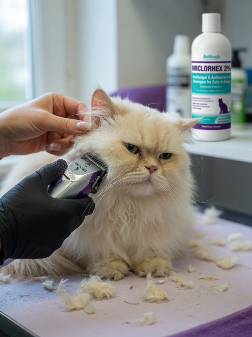
For localised infections in otherwise healthy animals, topical therapy alone may be sufficient. For cats and dogs with widespread disease, topical treatment is used alongside systemic antifungals.
Whole-Body Antifungal Rinses and Shampoos
Twice-weekly whole-body treatments with miconazole-chlorhexidine shampoo or lime sulphur dip are the most widely recommended approaches in current veterinary dermatology guidelines. Lime sulphur, whilst effective, carries a pungent odour and may stain surfaces, so it requires careful handling. Enilconazole rinse is also used in some regions and shows strong efficacy against M. canis.
Clipping the coat in long-haired animals reduces fungal load and improves penetration of topical agents. Clipped hair must be disposed of carefully, as it can remain infective in the environment for months.
Systemic Antifungal Therapy
Systemic treatment is recommended for multi-lesion infections, infections in immunocompromised animals, or in multi-pet households where rapid resolution is a priority.
- Itraconazole: The preferred oral antifungal in most current protocols. Administered at approximately 5 mg/kg daily or in pulse-dose regimens (one week on, one week off). It concentrates well in skin and hair follicles.
- Terbinafine: A useful alternative, particularly in cats, often used when itraconazole is not well tolerated. Dosed daily and generally well accepted.
- Griseofulvin: An older agent now largely superseded. Requires a fatty meal for absorption and carries teratogenic risk, meaning it must never be given to pregnant animals.
- Fluconazole: Less consistently effective against M. canis but used in some cases, particularly where cost is a consideration.
Duration of systemic therapy typically extends two weeks beyond clinical cure and two consecutive negative fungal cultures, which can mean six to twelve weeks of treatment in total. Stopping early is one of the most common causes of treatment failure and relapse.
Environmental Decontamination
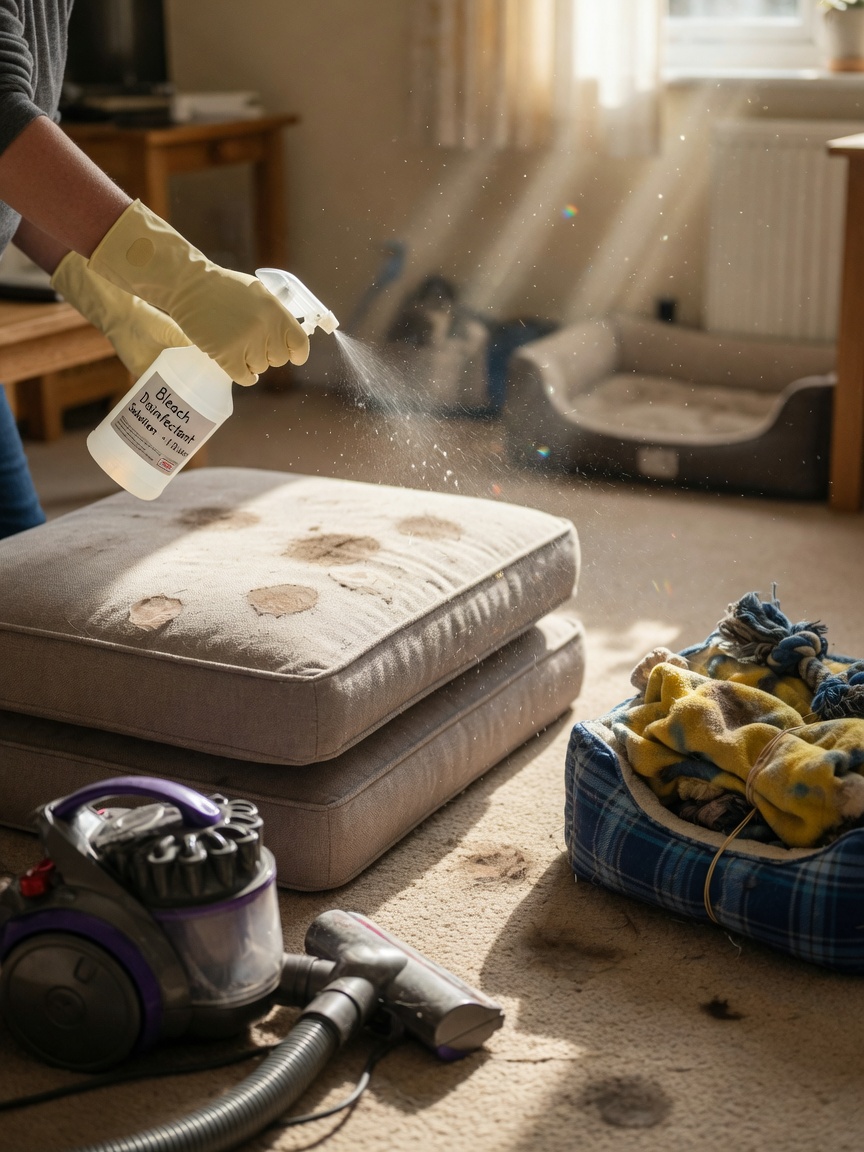
Treating the animal without addressing the environment is a recipe for reinfection. Dermatophyte spores can survive in carpets, bedding, grooming equipment, and furniture for 12–18 months under favourable conditions.
- Vacuum and discard the bag daily during active infection
- Disinfect hard surfaces with a diluted bleach solution (1:10) or an approved antifungal disinfectant
- Wash all bedding and soft furnishings at the highest safe temperature
- Discard or thoroughly disinfect grooming tools, collars, and toys
- Restrict the infected pet to easily cleaned areas where possible
Managing Multi-Pet and Zoonotic Risk
All in-contact animals should be screened, even if they show no signs. Asymptomatic carriers can sustain infection cycles within a household indefinitely. Children, elderly individuals, and immunocompromised people are at highest risk of contracting the infection from pets and should avoid direct contact with infected animals until treatment is well established.
Handwashing after handling affected pets is non-negotiable. If a household member develops circular, itchy skin lesions, they should consult a GP promptly and mention the animal's diagnosis.
Key Takeaways for Pet Owners
- Confirm diagnosis with fungal culture rather than relying on appearance alone
- Combine topical and systemic treatment for widespread or multi-pet infections
- Treat for the full recommended duration — two negative cultures before stopping
- Decontaminate the home environment throughout the treatment period
- Screen all in-contact animals, even those without visible lesions
- Protect vulnerable household members and seek medical advice if they develop skin changes
Dermatophytosis is manageable, but it demands consistency. Work closely with your veterinarian to confirm the diagnosis, select the appropriate protocol, and monitor treatment response with repeat cultures.