
When the Gut Leaks What the Body Needs Most
Protein is not simply a dietary nutrient — it is a structural and functional currency the body cannot operate without. Albumin, the most abundant protein in the bloodstream, maintains fluid balance, transports hormones and fatty acids, and sustains tissue repair. When the gut begins to leak protein faster than the body can replace it, the consequences reach far beyond the digestive system. Protein-losing enteropathy is one of the most serious gastrointestinal syndromes in dogs, and understanding it is essential for any owner facing this diagnosis.
What Is Protein-Losing Enteropathy?
Protein-losing enteropathy (PLE) is not a single disease but a syndrome — a pattern of protein loss through the gastrointestinal tract that results in dangerously low levels of circulating proteins, particularly albumin and globulins. In a healthy dog, the intestinal lining is selectively permeable: it allows digested nutrients to pass into the bloodstream while retaining proteins and preventing their escape into the gut lumen. When this barrier is disrupted — through inflammation, lymphatic obstruction, or structural damage — plasma proteins leak into the intestinal space and are lost in the faeces.
The resulting hypoalbuminaemia (low blood albumin) reduces the oncotic pressure that keeps fluid within blood vessels. Fluid migrates into body cavities and tissues, producing the clinical signs most commonly associated with advanced PLE: ascites (fluid in the abdomen), pleural effusion (fluid around the lungs), and peripheral oedema — particularly visible as swelling of the limbs or ventral abdomen.
Underlying Causes
PLE is always secondary to another condition that is disrupting intestinal integrity. Identifying that underlying cause is essential to selecting the right treatment.
Inflammatory Bowel Disease
IBD is among the most common causes of PLE. Severe, chronic intestinal inflammation disrupts the mucosal barrier and, in some forms, obstructs intestinal lymphatics, leading to combined protein and lymph leakage. Lymphoplasmacytic enteritis and eosinophilic enteritis are both associated with PLE when inflammation is extensive.
Intestinal Lymphangiectasia
This condition involves dilation and dysfunction of the intestinal lymphatic vessels, causing lymph — which is rich in proteins, fat-soluble vitamins, and lymphocytes — to leak directly into the gut. Lymphangiectasia may be primary (structural) or secondary to conditions that obstruct lymphatic drainage, including heart disease, neoplasia, or severe IBD. It produces a characteristic picture of PLE combined with low lymphocyte counts and very low cholesterol.
Other Causes
Intestinal neoplasia — particularly lymphoma — must always be considered and ruled out, as it can produce a PLE syndrome that superficially resembles inflammatory disease. Fungal infections (such as histoplasmosis in endemic regions), severe parasitism, and gastrointestinal ulceration are additional potential causes.
Breeds at Particular Risk
Soft-Coated Wheaten Terriers have a well-documented hereditary predisposition to a severe form of PLE, often combined with protein-losing nephropathy (protein loss through the kidneys). Yorkshire Terriers and Basenjis are also over-represented. However, PLE can occur in any breed, and the absence of a predisposed breed does not lower diagnostic suspicion when clinical signs are present.
Recognising and Diagnosing PLE
The clinical presentation of PLE varies with severity and the underlying cause. Early cases may show only subtle signs — intermittent soft stools, mild weight loss, reduced appetite. As protein levels fall, more dramatic signs emerge: a distended, fluid-filled abdomen, laboured breathing from pleural effusion, and limb swelling. Weight loss may be marked despite an adequate or even increased appetite. Some dogs present in a crisis state requiring emergency stabilisation.
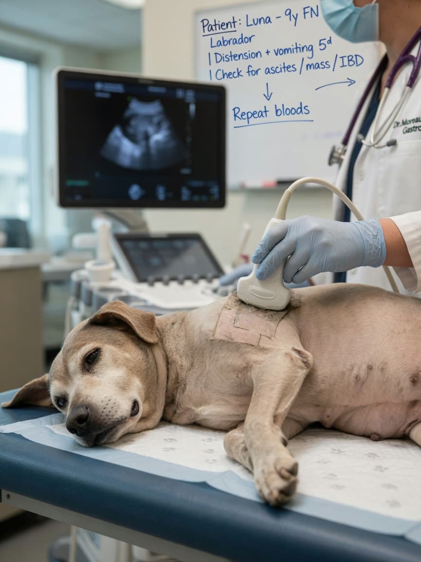
Blood work typically reveals hypoalbuminaemia and often hypo-globulinaemia, though the latter may be less pronounced when inflammatory disease is the driver. Cholesterol is frequently low. Lymphopenia (low lymphocyte count) is a hallmark of lymphangiectasia. Faecal alpha-1 proteinase inhibitor measurement can confirm intestinal protein loss when the diagnosis is uncertain. Abdominal ultrasound may show characteristic changes — mucosal striping, lymph vessel dilation, or lymph node abnormalities — that point towards the underlying cause. Intestinal biopsy remains essential for definitive diagnosis in most cases.
Treatment and Nutritional Management
Treatment targets both the underlying disease and the nutritional consequences of protein depletion.
Ultra-Low-Fat Dietary Therapy
Dietary fat restriction is critical in PLE, particularly when lymphangiectasia is involved. Dietary fat stimulates lymphatic flow; in a dog with dysfunctional or obstructed lymphatics, this perpetuates leakage. A diet containing less than 10% fat on a dry matter basis — often substantially lower — is typically required. Some dogs with severe lymphangiectasia require specially formulated ultra-low-fat diets or home-cooked meals under veterinary nutritionist supervision. Medium-chain triglycerides are sometimes included as an energy source, as they are absorbed directly into the portal blood rather than via the lymphatic system.
Protein Quality and Digestibility
A highly digestible, high-quality protein source supports the body's ability to replenish lost proteins. Novel or hydrolysed protein sources are used when dietary sensitivity is suspected as a contributing factor. Overall caloric density must be sufficient to support weight maintenance and protein synthesis without relying on fat as the primary energy source.
Medical Management
When IBD underlies PLE, immunosuppressive therapy is generally required. In severe cases with hypoalbuminaemia, plasma transfusions or synthetic colloids may be needed to provide short-term oncotic support whilst awaiting dietary and medical response. Vitamin supplementation — particularly fat-soluble vitamins and cobalamin — addresses predictable deficiencies. Anticoagulation is considered in some cases, as PLE increases the risk of thromboembolic complications.
Prognosis and Ongoing Management
PLE carries a guarded prognosis, and outcomes vary considerably depending on the underlying cause. Dogs with dietary-responsive or mild IBD-associated PLE may achieve long periods of remission with appropriate management. Those with lymphangiectasia often require lifelong dietary restriction and periodic monitoring. Lymphoma-associated PLE carries a more serious prognosis and requires oncological treatment.
- Monitor body weight and abdominal girth regularly at home
- Schedule frequent veterinary rechecks including albumin measurement, particularly in the first months of treatment
- Maintain strict dietary compliance — even occasional high-fat meals can trigger rapid clinical deterioration in dogs with lymphangiectasia
- Report any sudden worsening — increased abdominal distension, breathing difficulty, or collapse requires urgent veterinary attention
- Work closely with your vet or a veterinary internal medicine specialist; PLE management often benefits from specialist input