
When the Immune System Attacks the Skin
Pemphigus foliaceus is the most common autoimmune skin disease diagnosed in both dogs and cats. In a healthy animal, the immune system recognises and tolerates the body's own tissues. In pemphigus foliaceus, that tolerance breaks down. Autoantibodies — most commonly targeting desmoglein-1, a protein that holds skin cells together — cause separation within the superficial epidermis, producing the characteristic pustules and crusts that define this condition. It is not infectious, not contagious, and not caused by diet. It is, however, serious, and it requires accurate diagnosis and long-term veterinary management.
Who Is Most at Risk
Pemphigus foliaceus can affect dogs and cats of any breed, age, or sex, but certain patterns emerge in clinical practice.
In dogs, Akitas, Chow Chows, Dobermanns, Labrador Retrievers, and Bearded Collies appear to be predisposed, suggesting a heritable component to immune dysregulation. Middle-aged to older dogs are most commonly affected, though the disease can occur at any age. In cats, there is less clear breed predisposition, but Persian and Himalayan cats may be overrepresented in some reports.
The condition can arise spontaneously, or it may be triggered by prior drug exposure (drug-induced pemphigus), chronic skin disease, or prolonged ultraviolet light exposure. In a subset of cases, no identifiable trigger is found.
Clinical Signs: What to Look For
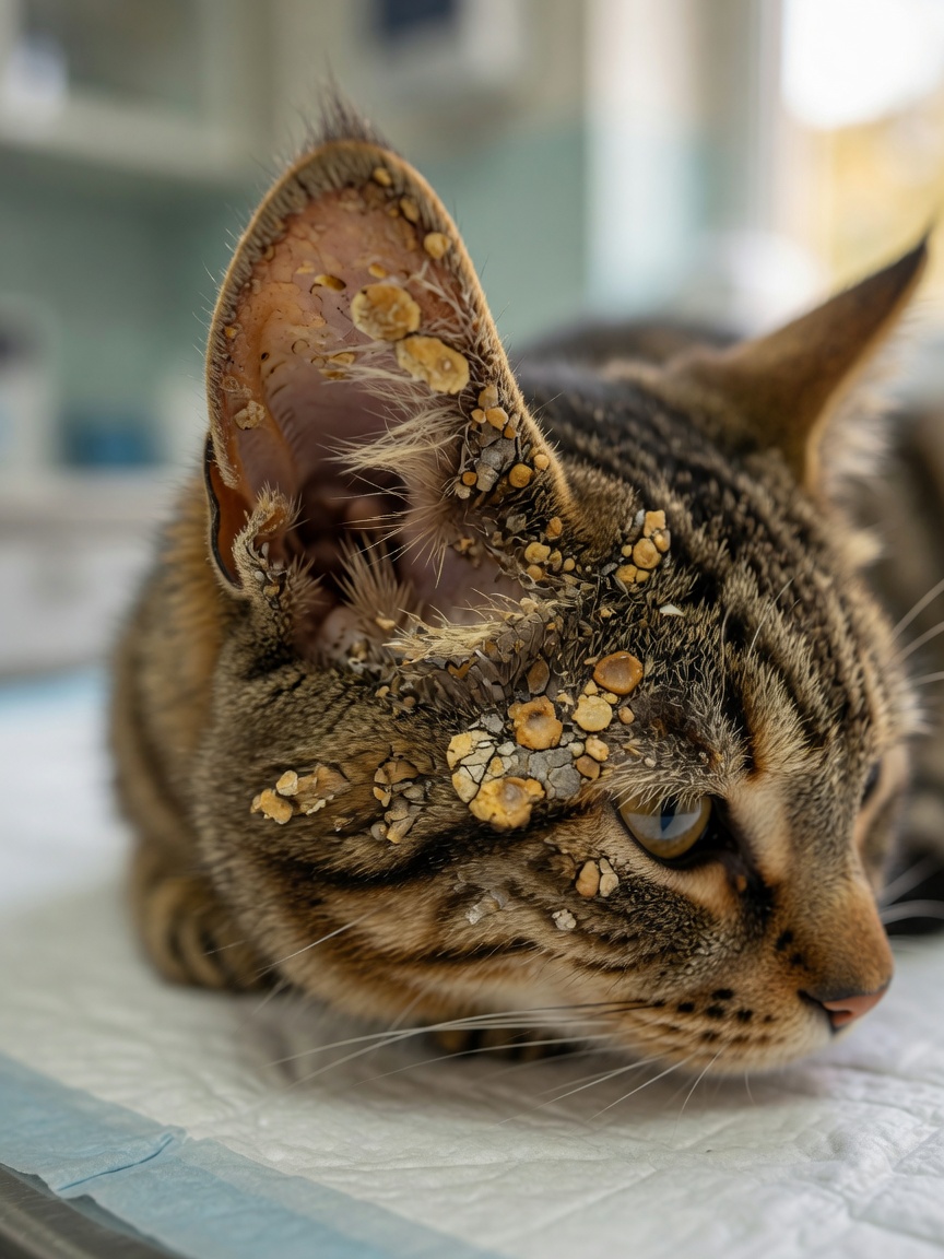
The lesion distribution in pemphigus foliaceus is distinctive and often helps clinicians narrow the differential diagnosis before biopsy results are available.
Primary Lesions
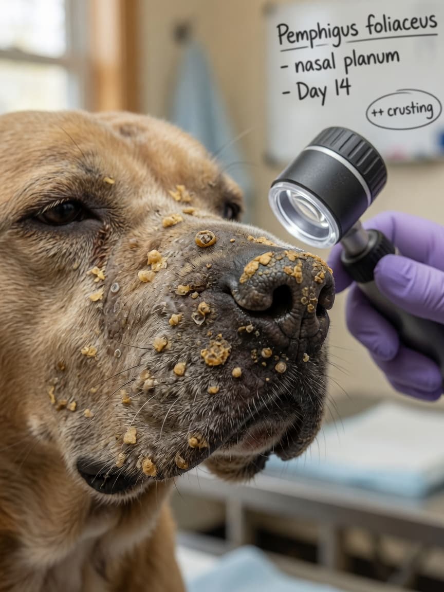
Pustules — sterile collections of neutrophils within the epidermis — are the primary lesion, but they rupture quickly and are often missed. What owners and clinicians typically observe are the secondary lesions: thick, adherent crusts; erosions; and collarettes (circular scale remnants left when pustules break).
Distribution Patterns
- Nasal planum and dorsal muzzle: almost universally affected in dogs
- Pinnae (ear flaps): a characteristic site, particularly in cats
- Periocular skin and footpads: common in dogs, with footpad involvement causing thickening and pain
- Nipples and nail folds: affected in some dogs
- Generalised truncal spread: occurs in more severely affected individuals
In cats, the head, neck, and pinnae are the most commonly affected areas. Footpad involvement is also well recognised in feline cases and can cause significant lameness.
Systemic signs — lethargy, fever, and inappetence — occur in more severely affected animals. Pruritus is variable; some animals are intensely itchy, others barely so.
Reaching a Diagnosis
Clinical presentation alone is suggestive but insufficient. Several other diseases mimic pemphigus foliaceus, including bacterial pyoderma, dermatophytosis, and other immune-mediated conditions. Confirmation requires skin biopsy.
Biopsies should be taken from intact pustules wherever possible, as these yield the most diagnostically useful tissue. If pustules have ruptured, early crusted lesions are the next best choice. Multiple samples from different sites improve diagnostic yield. Histopathology characteristically demonstrates subcorneal or intraepidermal pustules filled with neutrophils and acantholytic cells — detached, rounded epidermal cells that have lost their connections to neighbouring cells due to autoantibody activity.
Bacterial culture of pustule contents helps exclude infectious causes and guides antimicrobial selection if secondary infection is present. Direct immunofluorescence testing, detecting immunoglobulin deposits in the epidermis, can support the diagnosis but is not routinely required in all cases.
Immunosuppressive Treatment Protocols
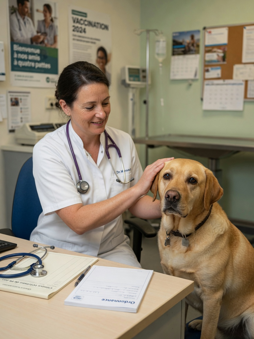
Because pemphigus foliaceus is driven by immune system activity, treatment centres on immunosuppression. The goal is to dampen autoantibody production and reduce epidermal inflammation sufficiently to allow skin healing, using the lowest effective dose to minimise long-term side effects.
Glucocorticoids
Prednisolone or methylprednisolone at immunosuppressive doses forms the foundation of initial therapy in most cases. Response is often rapid, with significant improvement within two to four weeks. Once remission is achieved, the dose is tapered gradually over months to find the minimum effective maintenance dose.
Steroid-Sparing Agents
Many animals require additional immunosuppressive drugs to achieve adequate control or to allow steroid tapering to safer long-term doses. Commonly used agents include:
- Azathioprine — widely used in dogs; requires monitoring of blood counts and liver enzymes as it can cause bone marrow suppression and hepatotoxicity
- Chlorambucil — preferred in cats, as azathioprine carries unacceptable toxicity risk in the feline species
- Ciclosporin — effective in some cases and increasingly used as a steroid-sparing option
- Mycophenolate mofetil — used in refractory cases
Long-Term Management and Monitoring
Pemphigus foliaceus is rarely cured. The majority of affected animals require lifelong or prolonged immunosuppressive therapy, and the disease may relapse even in animals that achieve complete remission. Regular veterinary monitoring is essential.
- Schedule blood counts and biochemistry panels at regular intervals, particularly for animals on azathioprine or chlorambucil
- Watch for signs of secondary infection — immunosuppressed skin is vulnerable to bacterial overgrowth
- Minimise sun exposure, particularly in dogs with nasal and facial lesions, as ultraviolet light can exacerbate disease
- Report any sudden worsening, new pustule crops, or systemic illness promptly
- Never adjust immunosuppressive doses without veterinary guidance — both under- and over-treatment carry significant risks
A diagnosis of pemphigus foliaceus is manageable with the right partnership between owner and veterinarian. Many dogs and cats achieve good quality of life on maintenance therapy, and some enter prolonged remission. Consistency, monitoring, and prompt attention to flares make the difference.