
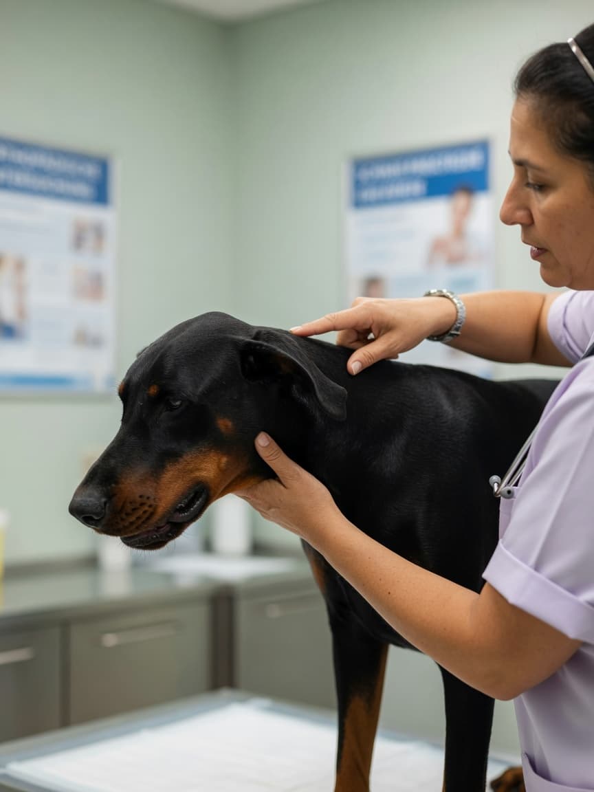
¿Qué es el Síndrome Wobbler?
El síndrome wobbler es el nombre común de la espondilopatía cervical (CSM) — una condición en la que las anomalías de las vértebras cervicales (cuello) comprimen la médula espinal o las raíces nerviosas en la región del cuello. La compresión interfiere con la transmisión de señales nerviosas entre el cerebro y las extremidades, produciendo la marcha característica inestable y tambaleante que le da nombre a la condición.
La CSM no es una enfermedad única y uniforme. Abarca una serie de anomalías estructurales que incluyen hernia discal intervertebral, inestabilidad de la columna vertebral, malformación ósea de las vértebras e hipertrofia (engrosamiento) de los ligamentos dentro del canal espinal. El resultado final en cada caso es el mismo: estrechamiento del canal espinal y compresión del delicado tejido en su interior.
Predisposiciones por Raza
El síndrome wobbler tiene una clara predilección por razas grandes y gigantes, con dos en particular que destacan:
Dobermann
El Dobermann es la raza más afectada por el síndrome wobbler, representando la mayoría de los casos en muchos estudios. En esta raza, la condición típicamente implica compresión asociada a disco, más frecuentemente en la unión C5-C6 o C6-C7. El inicio generalmente ocurre en la mediana edad, entre cinco y nueve años, aunque los perros más jóvenes pueden verse afectados. La condición en Dobermanns tiende a ser crónica y progresiva.
Gran Danés
Los Grandes Daneses desarrollan una forma de CSM que difiere del tipo Dobermann. Más comúnmente implica malformación ósea de las propias vértebras — a veces llamada el tipo "wobbler" o "disco" versus el tipo "óseo" — y tiende a afectar a perros más jóvenes, frecuentemente entre uno y tres años de edad. Otras razas gigantes como el Mastiff, San Bernardo y Galgo Irlandés también están representadas.
Signos Clínicos

La presentación del síndrome wobbler refleja la ubicación y severidad de la compresión de la médula espinal.
Déficits Propioceptivos y Marcha Tambaleante
El signo más característico es una marcha anormal en los miembros posteriores. Los perros afectados parecen tropezar o balancearse, colocando los pies de manera desigual y a veces cruzando los miembros posteriores mientras caminan. Esto ocurre porque la propiocepción — la conciencia sensorial de dónde están las extremidades en el espacio — está afectada por la compresión. Colocar la parte posterior del pie del perro en el suelo y observar qué tan rápido se corrige (la prueba de posicionamiento propioceptivo) a menudo revela déficits claros. Los miembros anteriores también pueden verse afectados, pero la ataxia de miembros posteriores es generalmente más pronunciada ya que los tractos de médula espinal relevantes son selectivamente vulnerables.
Dolor Cervical
Muchos perros con síndrome wobbler muestran signos de dolor de cuello. Pueden ser reacios a bajar la cabeza para comer o beber, resistir la flexión o extensión del cuello, o gritar cuando el cuello es palpado. Algunos perros adoptan una posición de cabeza característicamente rígida y baja. El dolor cervical puede ser intermitente y puede no siempre ser obvio en un único examen.
Afectación de Miembros Anteriores
Cuando la compresión es severa o afecta múltiples sitios, los miembros anteriores pueden mostrar debilidad, rigidez o zancada acortada — a veces descrita como una marcha de miembros anteriores "entrecortada". En los casos más severos, los perros pueden ser incapaces de caminar o pueden presentarse tetrapareticos (debilidad en los cuatro miembros).
Diagnóstico
Las radiografías simples de la columna cervical pueden sugerir el diagnóstico al revelar espaciamiento anormal entre vértebras, cambios óseos, o altura de disco reducida. Sin embargo, la resonancia magnética es el estándar de oro para definir la ubicación precisa, naturaleza y grado de compresión de la médula espinal. Proporciona el detalle necesario para la planificación quirúrgica e ayuda a identificar si uno o múltiples sitios están involucrados — una distinción importante que influye en la elección del tratamiento.
La mielografía (inyección de colorante de contraste en el líquido espinal para destacar el canal espinal bajo radiografía) fue la investigación históricamente elegida antes de que la resonancia magnética se volviera ampliamente disponible y aún se usa en algunas prácticas. La mielografía dinámica — imagenología del cuello en flexión y extensión — puede revelar compresión que solo está presente en ciertas posiciones.
Opciones de Tratamiento

Manejo Conservador
El manejo conservador tiene como objetivo reducir la inflamación y restringir el movimiento para permitir que la médula espinal se recupere de la compresión. Típicamente implica un período de reposo estricto, medicamento antiinflamatorio y alivio del dolor. Un collarín de cuello puede ser usado para limitar el movimiento cervical y reducir el estrés en los segmentos comprimidos.
El manejo conservador es más apropiado para perros con signos leves a moderados, aquellos considerados de riesgo quirúrgico muy alto debido a otras condiciones de salud, o propietarios que no pueden proseguir la intervención quirúrgica. Aproximadamente la mitad de los perros manejados conservadoramente muestran una mejora significativa, aunque la recurrencia y la progresión son comunes con el tiempo.
Opciones Quirúrgicas
La cirugía tiene como objetivo descomprimir la médula espinal y, cuando sea apropiado, estabilizar los segmentos vertebrales afectados. Se han descrito varios enfoques quirúrgicos, la elección depende de la naturaleza de la compresión, el número de sitios involucrados y la anatomía individual del perro.
El procedimiento de ranura ventral es una de las técnicas más comúnmente realizadas para la compresión asociada a disco. El cirujano aborda la columna cervical desde debajo del cuello, extirpa una pequeña ventana de material óseo y discal, y descomprime directamente la médula espinal. Es técnicamente exigente pero bien establecido, con buenos resultados reportados en muchas series.
Las técnicas de distracción-fusión se usan para estabilizar segmentos afectados mientras se alivia simultáneamente la compresión. Los implantes se colocan para separar las vértebras y mantener la corrección mientras ocurre la fusión ósea en las semanas y meses posteriores. Estos procedimientos han ganado favor para enfermedad de múltiples sitios y para casos donde la simple descompresión es improbable que sea suficiente.
Pronóstico
El pronóstico para el síndrome wobbler depende fuertemente de la severidad de los signos en la presentación, el número de sitios involucrados y el tratamiento perseguido. Los perros con signos leves a moderados que se someten a cirugía descompresiva exitosa tienden a hacer bien, con una proporción significativa mostrando mejora marcada. Los perros que no son ambulatorios en el momento de la cirugía tienen un pronóstico más reservado. La recurrencia en los mismos o sitios adyacentes es una complicación reconocida, particularmente en perros manejados a largo plazo.
La fisioterapia regular y la hidroterapia son adyuvantes valiosos tanto para el manejo quirúrgico como conservador, ayudando a mantener la masa muscular y la función propioceptiva durante el tratamiento y la recuperación.