
Two Tools, Two Very Different Purposes
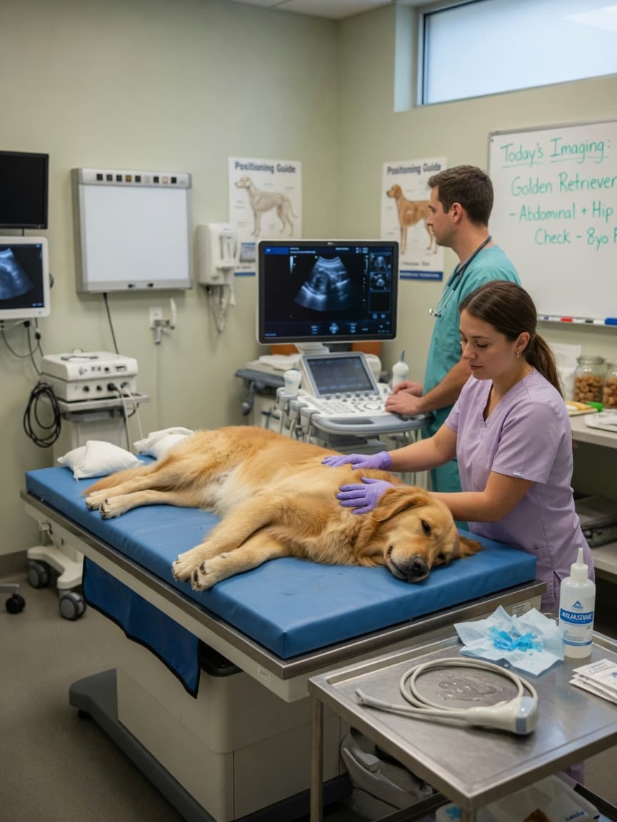
When your vet recommends imaging for your pet, it can be confusing when they suggest either an X-ray or an ultrasound — or sometimes both. These are not interchangeable options offering the same information at different price points. They work on entirely different physical principles and reveal different aspects of your pet's anatomy. Understanding the distinction helps you understand what your vet is looking for and why one modality suits a particular situation better than the other.
How X-Rays Work and What They Show
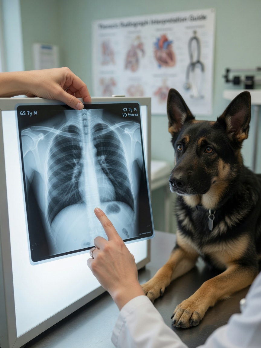
X-rays, or radiographs, pass a beam of radiation through the body. Different tissues absorb different amounts of radiation, and the result is a two-dimensional image showing density contrasts. Dense structures such as bone appear bright white because they absorb most of the beam. Air-filled spaces such as lungs appear dark. Soft tissues sit somewhere in between, appearing in shades of grey.
This makes X-rays excellent for assessing bony structures: fractures, joint disease such as arthritis or hip dysplasia, bone tumours, and spinal abnormalities all show clearly on radiographs. In the chest, X-rays are the primary tool for evaluating the lungs and heart size. Fluid in the lungs or chest cavity, pneumonia, tumours in lung tissue, and cardiac enlargement are all well-demonstrated on thoracic radiographs.
In the abdomen, X-rays are more limited because most of the organs are soft tissue with similar densities. However, they are excellent for identifying foreign bodies — a swallowed stone or bone will show clearly. They can reveal gas patterns in the intestines that indicate obstruction or bloat, assess overall organ size, and detect mineralised masses or bladder stones.
How Ultrasound Works and What It Reveals
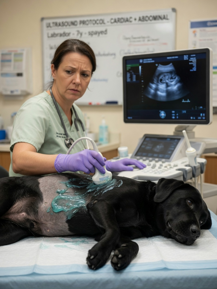
Ultrasound uses high-frequency sound waves, not radiation. A probe placed against the skin emits pulses of sound that bounce back differently from different tissue types. These returning echoes are processed in real time to produce a moving image on a screen.
Where X-rays show density, ultrasound shows texture and internal architecture. This makes it far superior for evaluating soft tissue organs in detail. The liver, spleen, kidneys, adrenal glands, bladder, and lymph nodes can all be examined for changes in their internal structure, nodules, masses, cysts, or abnormal blood flow using a technique called Doppler imaging.
Ultrasound is the imaging method of choice for abdominal investigations. It can detect a splenic mass missed on radiograph, characterise a kidney lesion, identify bladder wall thickening, or confirm a liver that appears enlarged on X-ray as containing multiple focal lesions rather than being diffusely diseased. These distinctions matter enormously for determining the correct next step.
Ultrasound is also the standard imaging tool in pregnancy assessment, allowing the vet to count foetuses, assess viability through detecting heartbeats, and estimate gestational age.
The Key Limitations of Each
X-rays cannot show the internal detail of soft tissue organs. A mass on a kidney or a nodule within the spleen may be completely invisible on a radiograph if it does not alter the organ's size or produce calcification. They are also a static, two-dimensional snapshot, which limits assessment of dynamic processes.
Ultrasound has its own constraints. Sound waves do not penetrate gas or bone. This means lungs — which are full of air — cannot be meaningfully imaged with ultrasound under normal circumstances. Bone blocks the sound entirely, making ultrasound useless for fractures or joint disease. Animals with a lot of intestinal gas can be challenging to image abdominally, and obesity can significantly reduce image quality.
When Both Are Used Together
It is not uncommon for your vet to recommend both modalities in the same visit, and this is not redundant. A dog presenting with unexplained weight loss might have chest X-rays taken to look for lung changes and heart size, plus abdominal ultrasound to examine the liver, spleen, and lymph nodes in detail. Together they provide a far more complete picture than either alone.
Similarly, a dog with a suspected abdominal mass might first have X-rays to assess the chest for spread and look for obvious abdominal changes, followed by ultrasound to characterise the mass in detail and guide a fine needle aspirate or biopsy.
Sedation and Preparation
X-rays often require sedation or even general anaesthesia in anxious, painful, or uncooperative animals, particularly when precise positioning is needed. Thoracic radiographs for cardiac assessment need to be taken at full inspiration, which requires a cooperative patient.
Ultrasound is generally well tolerated in calm patients without sedation, though anxious animals may need light sedation to allow adequate contact and imaging time. For abdominal ultrasound, an overnight fast is ideal to reduce intestinal gas, and the fur over the imaging area is shaved to allow the probe to make direct contact with the skin.
The Role of the Person Operating the Equipment
Regardless of which modality is used, the quality of information gained is heavily dependent on the skill of the operator. Ultrasound in particular is highly operator-dependent — the same patient can yield very different information depending on who is holding the probe and interpreting what they see. In complex cases, referral to a specialist in veterinary diagnostic imaging for an expert scan is well worth considering, and many first-opinion practices offer referral to visiting imaging specialists or internal medicine specialists.
Both tools are genuinely valuable. Neither is universally superior — they are simply suited to different questions. Knowing which question needs answering is how your vet decides which tool to reach for.