

What Is Vestibular Disease in Dogs?
The vestibular system is the body's balance and spatial orientation network, comprising receptors in the inner ear and pathways running through the brainstem to the cerebellum. When this system malfunctions, the result is a sudden loss of balance that can appear alarming — and to many owners, frightening enough to resemble a stroke. In reality, most dogs presenting with acute vestibular signs have idiopathic vestibular disease: a self-limiting condition with an excellent prognosis that typically resolves without any specific treatment.
Vestibular disease is one of the most commonly misunderstood conditions in dog medicine. The dramatic onset, the distressing symptoms, and the dog's apparent suffering can convince owners that something catastrophic has happened. Knowing what to look for — and what the signs actually mean — can spare both you and your dog unnecessary anxiety, while also ensuring you know when more urgent investigation is genuinely warranted.
Signs and Symptoms: What Vestibular Disease Looks Like
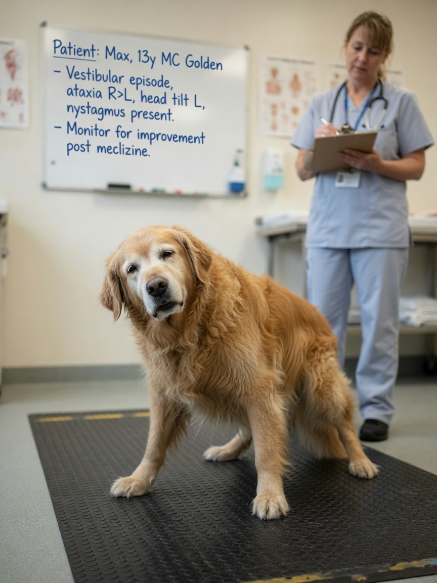
Idiopathic vestibular disease (also called old dog vestibular disease, because it most commonly affects dogs over eight years of age) comes on rapidly — often within hours and sometimes apparently overnight. The classic signs include:
- Head tilt: The dog holds its head persistently tilted to one side (toward the affected ear). This is one of the most consistent and recognisable signs.
- Nystagmus: Rapid, involuntary flickering of the eyes. In vestibular disease, this is typically horizontal (eyes flicker side to side) or rotatory (eyes rotate in a circular direction). The fast phase of the nystagmus usually beats away from the side of the lesion.
- Ataxia: The dog staggers, stumbles, and loses the ability to walk in a straight line. Many dogs circle or drift towards the affected side.
- Rolling: In severe cases, the dog cannot right itself and rolls continuously. This is terrifying to witness but is a feature of peripheral vestibular dysfunction, not a sign that the brain itself is failing.
- Vomiting and nausea: The dizziness caused by vestibular dysfunction is profoundly nauseating, comparable to severe motion sickness in people. Many affected dogs vomit repeatedly, particularly in the first 24–48 hours.
- Reluctance to eat or drink: A consequence of nausea and spatial disorientation.
Dogs with vestibular disease remain alert and responsive throughout — they are aware of what is happening around them even if they cannot coordinate their movements normally. Consciousness is entirely preserved.
Idiopathic Old Dog Vestibular Disease: What Causes It?
The honest answer is that in the idiopathic form — which accounts for the majority of acute vestibular presentations in older dogs — the precise cause is unknown. The working hypothesis is that it involves a transient disruption to the peripheral vestibular apparatus in the inner ear, but no definitive pathological mechanism has been identified. It is not caused by a stroke, a tumour, or an infection in most cases, though all of these can cause similar signs and must be considered.
Other causes of vestibular syndrome that must be ruled out include:
- Otitis media/interna (middle or inner ear infection)
- Hypothyroidism (which can cause peripheral vestibular neuropathy)
- Nasopharyngeal polyps
- Ototoxic drug exposure (certain antibiotics, particularly aminoglycosides)
- Central lesions (brainstem or cerebellum tumours, infarcts, or inflammatory disease)
A thorough clinical examination, otoscopy (inspection of the ear canal), basic blood tests, and thyroid function testing will identify or exclude most non-idiopathic causes without the need for advanced imaging in straightforward presentations.
Recovery Timeline: What to Expect
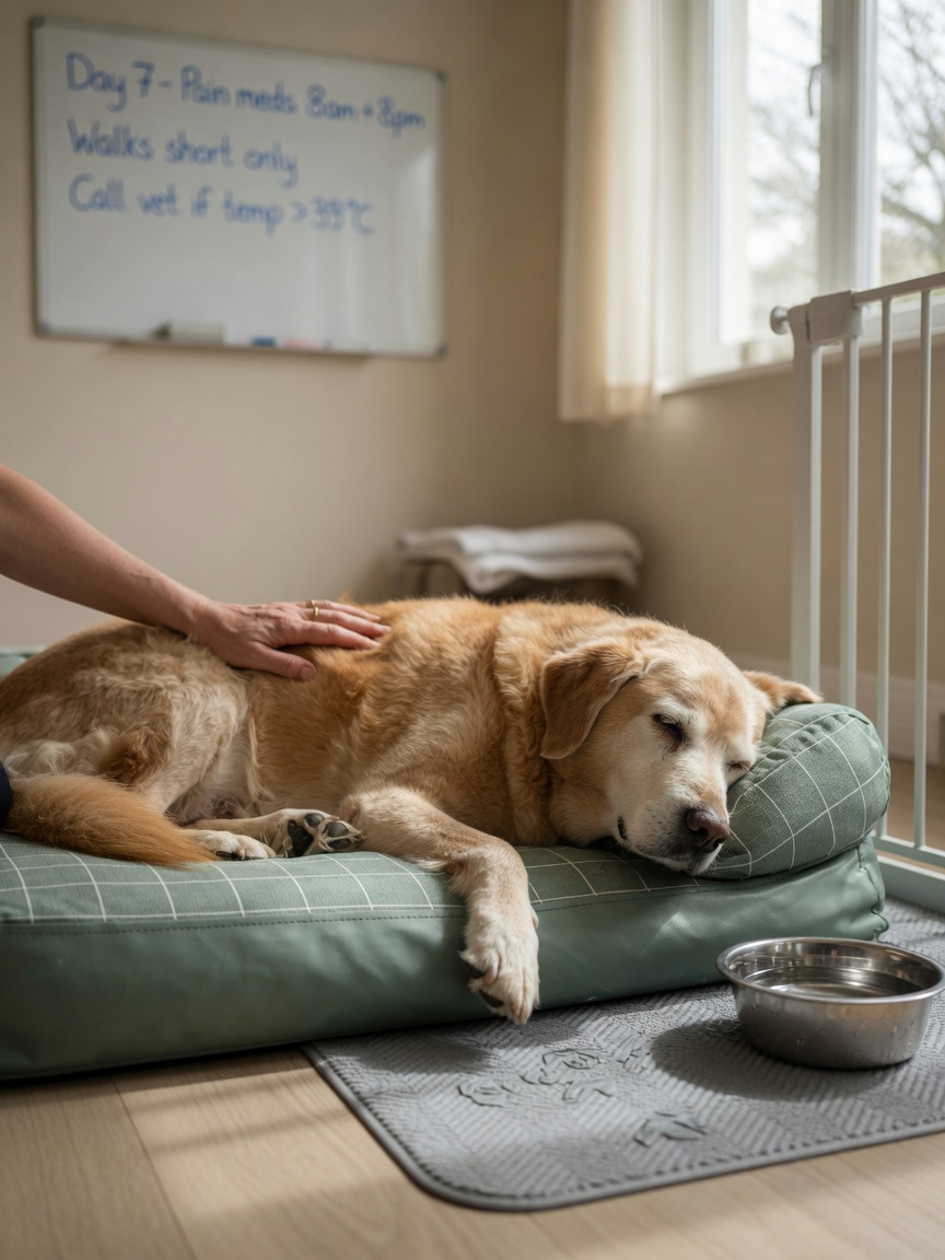
The natural history of idiopathic vestibular disease is reassuringly positive. Most dogs begin to show meaningful improvement within 72 hours, and the majority make a full or near-full recovery within two to four weeks. In some dogs, a mild residual head tilt persists long-term, but this has no impact on their quality of life or day-to-day functioning.
The first 24–48 hours are usually the worst. Dogs in this phase are often unable to stand, may be rolling, and are frequently vomiting. This is when the condition is most frightening to owners and when the resemblance to a stroke is strongest. But the trajectory is typically one of steady improvement rather than deterioration: nystagmus tends to reduce first, followed by improving coordination, with the head tilt resolving last.
During recovery, practical nursing care makes a significant difference:
- Confine the dog to a small, safe space where it cannot injure itself by falling
- Provide a non-slip surface for walking, as ataxia worsens on slippery floors
- Offer water little and often, as nausea often suppresses thirst
- Assist with outdoor toileting, supporting the dog if needed
- Avoid car travel, which will worsen motion sickness
Anti-Nausea Treatment: Cerenia and Supportive Care
While most dogs recover without specific treatment for the underlying condition, anti-nausea medication is an important and effective tool for improving comfort during the acute phase. Maropitant (Cerenia) is the most commonly used antiemetic in veterinary practice and is highly effective at controlling the vomiting and nausea associated with vestibular disease. It can be given by injection at the vet clinic or prescribed as tablets for home administration.
Meclizine, an antihistamine with vestibular-suppressant properties, is used in some practices as an additional measure. Sedatives are occasionally used in dogs that are extremely distressed or injuring themselves through rolling, though these should be used cautiously as they can mask signs that might indicate neurological deterioration.
Intravenous fluid support may be required for dogs that cannot keep food or water down, particularly in elderly patients where dehydration is a real concern over the first couple of days.
Distinguishing Vestibular Disease from a Brainstem Stroke
This distinction is clinically critical. Peripheral vestibular disease (affecting the inner ear) carries an excellent prognosis. Central vestibular disease (affecting the brainstem or cerebellum) may be caused by infarction, haemorrhage, or a mass lesion, and carries a much more variable prognosis requiring urgent investigation and different management entirely.
Key clinical features that suggest central rather than peripheral disease include:
- Vertical nystagmus: Eyes flickering up and down rather than side to side or rotationally. This pattern is almost always a sign of central vestibular dysfunction and should prompt urgent neurological investigation.
- Nystagmus that changes direction depending on head position
- Absence of improvement in head tilt: In peripheral disease, the head tilt typically begins to improve within days. A head tilt that does not improve at all by 72 hours is more suspicious for a central lesion.
- Altered consciousness or mentation: Depression, stupor, or unusual behaviour suggests brainstem involvement
- Cranial nerve deficits: Facial paralysis, altered swallowing, or drooping of the lip alongside vestibular signs
- Proprioceptive deficits: Knuckling or stumbling of all four limbs rather than just unsteadiness
If any of these features are present, MRI of the brain is strongly recommended and should not be delayed.
Cavalier King Charles Spaniels and Vestibular Disease
Cavalier King Charles Spaniels (CKCS) have an increased prevalence of vestibular disease, including both peripheral and central forms. They are also predisposed to syringomyelia and Chiari-like malformation, conditions which can sometimes present with neurological signs that overlap with vestibular disease. Any CKCS presenting with vestibular signs that do not follow the expected trajectory of rapid improvement should be referred promptly for specialist neurological assessment and MRI, rather than being assumed to have the straightforward idiopathic form of the condition.
Written by Julia Wrenfield