

The Goose Honk That Owners Never Forget
There is a particular cough that vets describe as sounding exactly like a goose honking — harsh, paroxysmal, and often triggered by excitement, exercise, or even just pulling on a lead. If you own a Yorkshire Terrier, Pomeranian, Chihuahua, or Pug and this description is immediately familiar, you have likely already encountered tracheal collapse. It is one of the most common respiratory conditions in small and toy breeds, and whilst it is manageable in most cases, it does require proper understanding and long-term commitment from owners.
What Happens to the Trachea
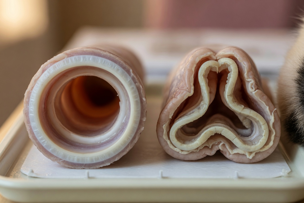
The trachea — the windpipe — is normally held open by a series of C-shaped cartilage rings. In dogs with tracheal collapse, these rings lose their rigidity and flatten, causing the tracheal walls to cave inward during breathing. The result is a narrowed, partially obstructed airway that produces the characteristic cough and, in more severe cases, genuine respiratory distress.
The collapse can occur in the cervical (neck) portion of the trachea, which tends to collapse on inhalation, or the thoracic (chest) portion, which tends to collapse on exhalation. Many affected dogs have involvement of both regions. In advanced cases, the condition may be complicated by concurrent collapse of the bronchi (bronchomalacia) and involvement of the larynx.
Why Small Breeds Are Disproportionately Affected
Genetic Cartilage Weakness
The primary reason is a genetically determined weakness in the composition of the tracheal cartilage. In affected dogs, the cartilage rings have reduced glycosaminoglycan and chondroitin content and increased water content, making them structurally compromised. This is an inherent characteristic of many small and toy breeds — it is not caused by anything the owner has done. Yorkshire Terriers, Pomeranians, Chihuahuas, Toy and Miniature Poodles, and Pugs are among the most commonly affected breeds, though the condition can occur in any small dog.
Brachycephalic Complications
In brachycephalic breeds such as Pugs, French Bulldogs, and Shih Tzus, tracheal collapse often coexists with brachycephalic obstructive airway syndrome (BOAS) — a cluster of anatomical abnormalities including narrowed nostrils, elongated soft palate, and narrowed trachea at baseline. The increased respiratory effort these dogs exert to overcome their airway resistance places additional stress on the tracheal cartilage over time.
Recognising the Signs and Grading Severity
The classic presentation is a paroxysmal, dry, honking cough, often worse with excitement, exercise, eating or drinking, heat and humidity, or tracheal pressure from a collar. Episodes may end in gagging or retching. Most dogs with mild to moderate disease are otherwise bright and comfortable between episodes.
In more severe disease, signs escalate. Dogs may show persistent respiratory effort, cyanosis (blue-tinged gums indicating oxygen deficit), weakness, or collapse. These are emergency presentations requiring immediate veterinary attention.
Tracheal collapse is graded from I to IV based on the degree of narrowing observed during diagnostic examination, with Grade I representing mild flattening and Grade IV representing complete collapse. Grade does not always directly predict clinical severity — some dogs with Grade IV collapse are more stable than expected, whilst Grade II dogs with significant concurrent disease may be highly symptomatic.
Diagnosis
Diagnosis combines signalment, clinical history, and imaging. Thoracic and neck radiographs can demonstrate tracheal narrowing, though they capture only a moment in the respiratory cycle and may miss dynamic collapse. Fluoroscopy — real-time moving X-ray — is more sensitive for detecting collapse at different phases of breathing. Tracheoscopy (endoscopic visualisation of the trachea under anaesthesia) is the most accurate method and allows direct grading, but carries some risk in compromised patients. CT scanning provides excellent anatomical detail and is increasingly used at specialist centres. Concurrent conditions including left-sided heart enlargement (which can press on the left main bronchus), obesity, and dental disease should also be assessed as they can worsen clinical signs.
Management: Medical and Surgical
Medical Management
The majority of dogs with tracheal collapse — particularly those with Grade I to III disease — are managed medically. The approach is multifaceted. Cough suppressants (antitussives) reduce the self-perpetuating cycle in which coughing irritates the trachea, provoking further coughing. Bronchodilators help by reducing bronchospasm and improving airflow. Anti-inflammatory doses of corticosteroids are used short-term during acute exacerbations to reduce airway inflammation and swelling. Sedatives such as butorphanol or acepromazine are invaluable during severe episodes to reduce respiratory effort and anxiety.
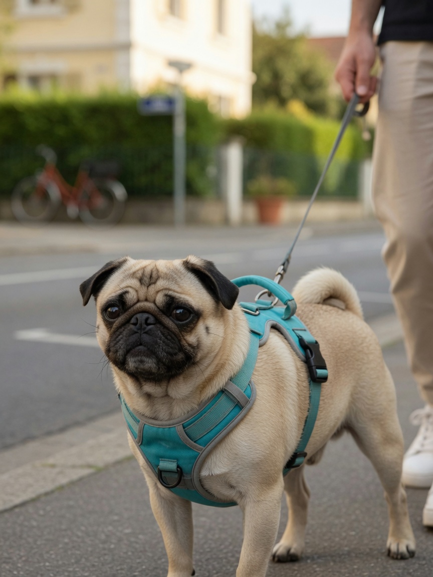
Weight management is one of the single most impactful interventions available. Even modest excess weight significantly worsens respiratory effort and clinical signs; achieving and maintaining ideal body condition is a genuine priority rather than an optional extra. Collar replacement with a harness removes pressure from the trachea during walks. Avoiding exposure to smoke, aerosols, and dusty environments reduces airway irritation.
Surgical Options
For dogs that fail to respond adequately to medical management, or for younger dogs with severe disease, surgical intervention may be considered. Extraluminal tracheal ring prostheses — rigid rings placed around the outside of the trachea to provide structural support — have been used with reasonable success rates for cervical tracheal collapse, though they carry risks including laryngeal paralysis. Intraluminal tracheal stenting, placing a self-expanding metal stent inside the trachea, is an endoscopic procedure increasingly used in specialist settings, particularly for thoracic tracheal collapse and in dogs where surgery is not feasible. Stents are effective but carry long-term complications including stent fracture, migration, and progressive collapse above or below the stent.
Living Well With Tracheal Collapse
- Always use a harness rather than a neck collar on an affected dog — even gentle lead pressure can trigger coughing.
- Keep your dog at an ideal body weight; obesity is one of the most controllable factors worsening this condition.
- Avoid exposing your dog to smoke, aerosols, air fresheners, or dusty environments.
- Keep excitement and strenuous exercise within manageable limits — gradual, low-intensity activity is preferable to bursts.
- Have a plan in place with your vet for acute episodes — knowing when to seek emergency care could save your dog's life.
- Work closely with your veterinarian to tailor a medical management plan; do not adjust medications without guidance.
- Discuss referral to a specialist if medical management is not achieving adequate control.