
That Little Hammer Does a Lot More Than You Think
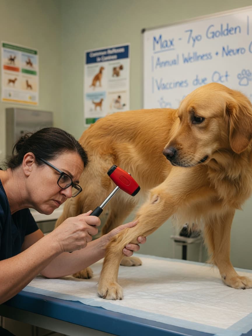
If you have ever watched a vet tap your dog's knee with a small rubber hammer and then nod thoughtfully, you may have wondered what they were actually measuring. That brief, automatic kick of the leg is called the patellar reflex, and it is one of the most reliable windows vets have into the health of your dog's nervous system. A single reflex test can help localise a lesion to a specific section of the spinal cord — without a single scan.
How the Patellar Reflex Works
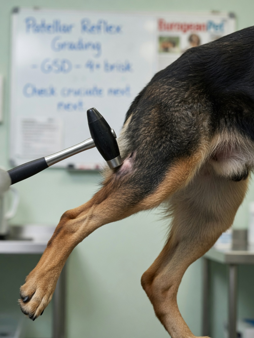
The patellar reflex is a simple arc. When the patellar tendon is tapped, sensory fibres in the quadriceps muscle send a signal up into the spinal cord at the lumbar segments — specifically L4 to L6 in dogs. The signal crosses directly to motor neurones that fire back down to the same muscle, producing the characteristic forward kick. Crucially, the brain is not involved. This is a local, spinal cord reflex, which is precisely what makes it so diagnostically useful.
Vets assess three qualities: whether the reflex is present, whether it is reduced (hyporeflexia), or whether it is exaggerated (hyperreflexia). Each finding points in a different neurological direction.
What Different Reflex Findings Tell the Vet
Absent or Reduced Patellar Reflex
A weak or absent response indicates a lower motor neurone problem. This means damage is at or near the reflex arc itself — the L4–L6 spinal cord segments, the nerve roots exiting there, or the femoral nerve running to the quadriceps. Common causes include intervertebral disc disease at the lumbar level, a tumour pressing on nerve roots, trauma, or peripheral nerve disease. The affected leg will often look flaccid and muscles may begin to waste relatively quickly.
Exaggerated Patellar Reflex
An overactive response points in the opposite direction: upper motor neurone disease. This means the problem sits above the reflex arc — typically in the thoracic spinal cord or even higher. The descending pathways that normally modulate and dampen reflex responses are disrupted, so the reflex fires without its usual brakes. Dogs with a thoracolumbar disc herniation, for example, may show exaggerated patellar reflexes in the hind limbs alongside hindlimb weakness or paralysis.
The Full Neurological Examination
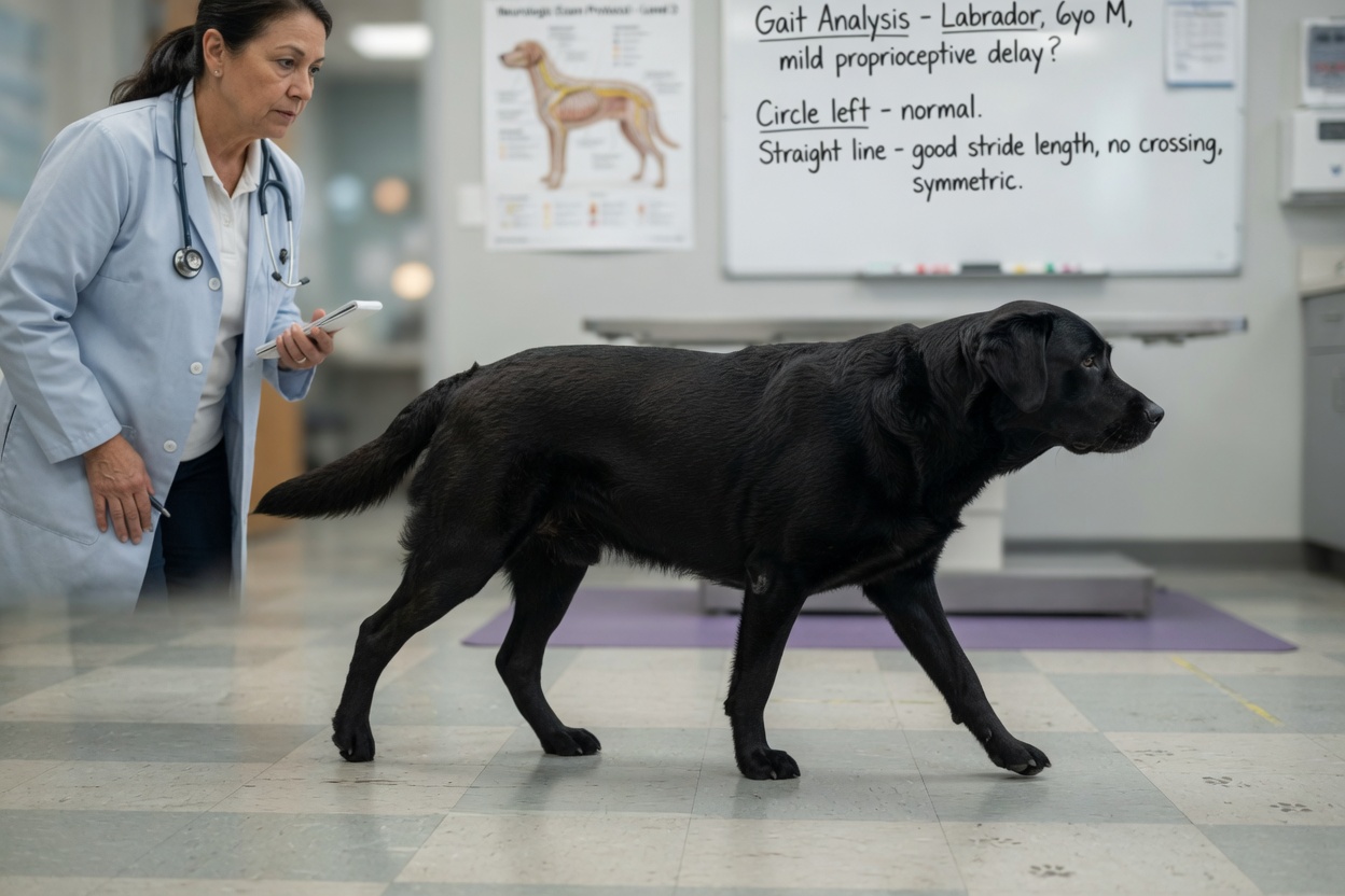
The patellar reflex is just one component of a structured neurological exam. A thorough assessment follows a logical sequence designed to answer one central question: where in the nervous system is the problem?
Mentation and Posture
The vet will observe your dog the moment it enters the room. Is it alert and responsive, or dull and confused? Is it holding its head at an angle? These observations can immediately suggest forebrain, brainstem, or vestibular involvement before a single test is performed.
Gait Analysis
Watching the dog walk in a straight line, turn in circles, and navigate a small obstacle reveals ataxia (uncoordinated movement), paresis (weakness), or paralysis. The pattern of limb involvement — all four limbs, just the back two, or one side — helps narrow the location of the lesion considerably.
Postural Reactions
Tests such as hopping, wheelbarrowing, and proprioceptive placing (placing the foot dorsal-side down and watching the dog correct it) assess whether the brain and spinal cord are communicating effectively. These are often the first responses to deteriorate when neurological disease sets in, even before obvious weakness appears.
Spinal Reflexes
Beyond the patellar reflex, vets assess the withdrawal reflex in all four limbs, the perineal reflex, and the panniculus reflex — a twitch of the skin muscle when the spine is lightly stimulated. Each reflex tests a different level of the spinal cord and helps build a neurological map.
Cranial Nerve Assessment
The twelve cranial nerves govern everything from eye movement and facial sensation to swallowing and tongue control. Vets check pupil symmetry, the menace response, eye position, jaw tone, and the gag reflex. Deficits here point to brainstem or peripheral cranial nerve lesions rather than spinal cord disease.
Why Localisation Matters Before Imaging
Before reaching for an MRI or CT scan, a skilled neurologist can often localise a lesion to within one or two spinal cord segments using the neurological exam alone. This matters for two reasons. First, it directs imaging — there is little value in scanning the neck if the exam points to the lumbar spine. Second, in emergency situations where scanning is not immediately available, the neurological exam guides treatment decisions and urgency.
The exam also provides a baseline. Serial neurological assessments over hours or days can reveal whether a patient is deteriorating, stable, or improving — information that influences whether surgery is needed and how urgently.
When to See a Vet
Any dog showing sudden hindlimb weakness, dragging a limb, appearing to lose coordination, or vocalising when touched along the spine warrants prompt veterinary attention. These can be signs of acute spinal cord compression, which may be reversible with early intervention. Do not wait to see if things improve on their own.
Dogs with chronic, slowly progressive weakness — particularly large and giant breeds — should also be assessed, as degenerative conditions of the spine and nervous system are common and manageable when caught early.
Key Points to Remember
- The patellar reflex tests a specific arc in the L4–L6 spinal cord segments
- A reduced reflex suggests lower motor neurone disease; an exaggerated one suggests upper motor neurone disease
- A full neurological exam includes gait, posture, postural reactions, spinal reflexes, and cranial nerve function
- Localisation guides imaging and treatment decisions before any scan is taken
- Sudden limb weakness or incoordination in your dog requires same-day veterinary assessment