
When One Eye Tells the Whole Story
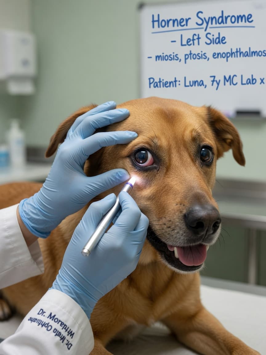
Imagine noticing one morning that your dog's eyelid is drooping, the eye itself looks slightly sunken, the pupil is smaller than the other side, and the third eyelid has crept across the inner corner. You have not seen any trauma. Your dog seems otherwise fine. What you are likely looking at is Horner syndrome — and that cluster of four subtle signs can point to a lesion anywhere along a long and winding nerve pathway that runs from the brain, down the spinal cord, out through the chest, up the neck, and into the eye.
The Anatomy Behind the Signs
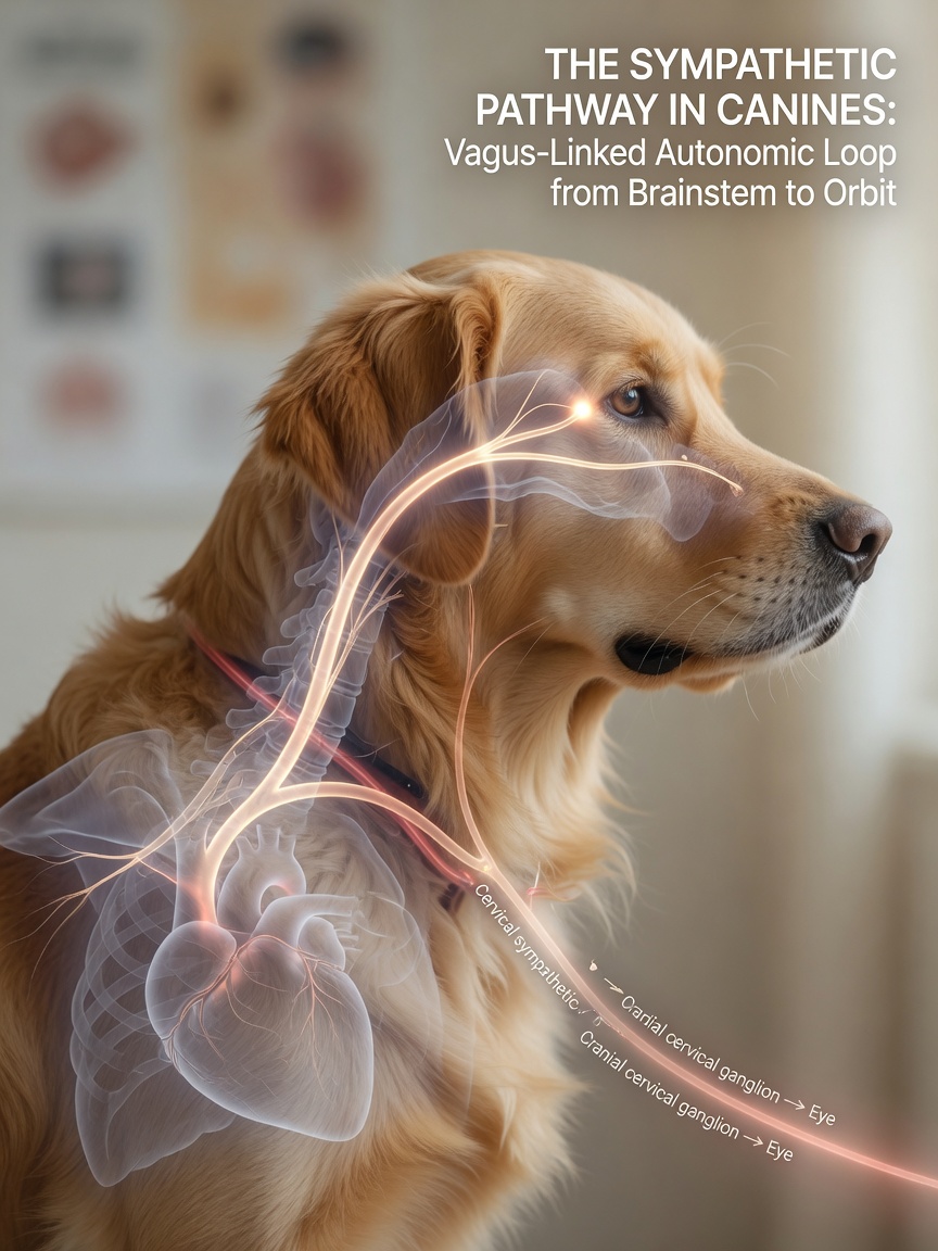
Horner syndrome results from disruption of the sympathetic nerve supply to the eye and surrounding structures. The sympathetic pathway to the eye is unusually long. It originates in the hypothalamus, descends through the brainstem and cervical spinal cord, exits at the thoracic spine, loops around structures in the chest including the heart base and the lung apex, travels back up the neck alongside the carotid artery, and finally reaches the eye via branches that run with the trigeminal nerve.
When this pathway is interrupted at any point, the same four signs appear: ptosis (drooping upper eyelid), miosis (constricted pupil), enophthalmos (sunken appearance of the eye), and protrusion of the third eyelid. The side affected always corresponds to the side of the lesion.
Classifying the Location: First, Second, and Third Order
Neurologists classify Horner syndrome by which part of the three-neurone pathway is damaged, and this classification guides the diagnostic workup significantly.
First-Order Horner Syndrome
The lesion involves the pathway from the hypothalamus to the thoracic spinal cord. Causes include cervical spinal cord tumours, disc disease in the neck or upper thoracic region, or infarcts. These patients typically have additional neurological signs such as weakness or ataxia in the limbs.
Second-Order Horner Syndrome
The lesion is in the chest — affecting the nerve roots at T1–T3 or the sympathetic trunk in the thorax. Causes include brachial plexus avulsion (a stretch injury to the nerves of the forelimb, commonly from being hit by a car), thoracic masses, or mediastinal lymphoma. This is the form most commonly seen in cats. In brachial plexus avulsion, the forelimb on the same side will also show weakness or paralysis.
Third-Order Horner Syndrome
The lesion affects the pathway from the cranial thorax up through the neck and into the orbit. This is the most common form in dogs and is frequently idiopathic — meaning no cause is ever found. Causes when identified include otitis media or interna (middle or inner ear infection), retrobulbar masses, trauma to the neck, or carotid artery disease.
How Vets Investigate Horner Syndrome
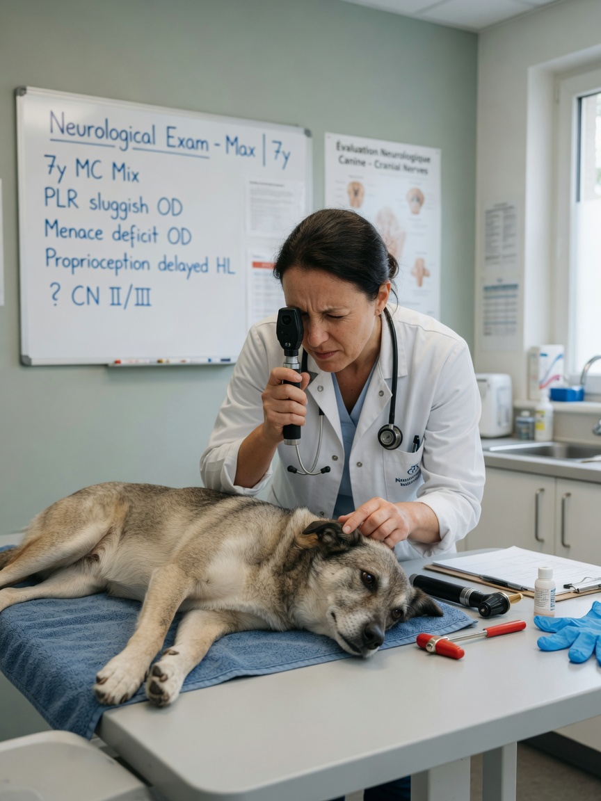
The first step is a full neurological and physical examination to look for additional signs that indicate where in the pathway the problem lies. A dog with Horner syndrome plus hindlimb ataxia is a very different case from one with Horner syndrome plus a head tilt and vestibular signs, or one with Horner syndrome as the only abnormality.
Pharmacological testing using dilute apraclonidine or phenylephrine eye drops can help differentiate pre-ganglionic from post-ganglionic lesions, though this test is more commonly employed in specialist neurology settings.
Depending on the clinical picture, investigations may include otoscopy and ear flushing under anaesthesia, thoracic radiographs, neck and chest ultrasound, MRI of the brain, neck, or thorax, and haematology and biochemistry to look for systemic disease.
Horner Syndrome in Cats
In cats, second-order Horner syndrome is disproportionately common. The thoracic pathway in cats is particularly vulnerable to mediastinal masses — lymphoma is the most frequent culprit — and to bite wounds over the neck and thorax. Any cat presenting with Horner syndrome should have thoracic radiographs taken promptly even if chest signs are not obvious. A large mediastinal mass can be present with minimal respiratory symptoms in early stages.
Idiopathic Horner syndrome does also occur in cats, though it is less common than in dogs.
Prognosis and Recovery
Prognosis depends entirely on the underlying cause. Idiopathic third-order Horner syndrome in dogs carries an excellent prognosis — the majority resolve spontaneously within six to eight weeks, though some take longer and a small number persist indefinitely without causing the animal any discomfort. The signs are cosmetically noticeable but do not impair vision or quality of life.
Horner syndrome secondary to otitis media often resolves once the ear infection is treated, though it can take weeks to months for sympathetic function to return. Horner syndrome associated with brachial plexus avulsion carries a much more guarded prognosis, particularly for the limb function, with the eye signs again causing no direct discomfort.
What You Should Do
- Consult a vet promptly if you notice the four classic signs — drooping lid, small pupil, sunken eye, visible third eyelid — on one side of your pet's face
- Do not assume it is cosmetic; the cause may require urgent treatment
- Bring any history of recent trauma, ear problems, or respiratory signs to the vet's attention
- Be prepared for imaging — identifying the location of the lesion often requires radiographs or MRI
- If diagnosed as idiopathic, monitor for resolution and report any new neurological signs immediately