
The Silent Thief of Sight — Until It Is Not
Glaucoma is often described as a silent disease in humans, where pressure builds gradually and painlessly over years. In dogs, the reality is frequently far more dramatic. Acute canine glaucoma is an emergency — a rapidly escalating rise in pressure inside the eye that causes significant pain and can destroy vision within 24 to 72 hours. Despite this urgency, it is commonly misread as conjunctivitis or a general eye irritation, costing dogs the sight they might otherwise have kept.
Understanding Intraocular Pressure
The eye continuously produces a fluid called aqueous humour, which flows through the anterior chamber and drains out via a structure called the iridocorneal angle. This drainage maintains a stable intraocular pressure — normal in dogs is roughly 10 to 25 mmHg. When drainage is obstructed or reduced, fluid accumulates and pressure rises. Elevated pressure damages the optic nerve and retinal cells, causing irreversible vision loss. The faster and higher the pressure rises, the more rapid and severe the damage.
Primary Versus Secondary Glaucoma
Primary Glaucoma
Primary glaucoma results from an inherited structural abnormality in the drainage angle and is not associated with another underlying disease. It tends to affect certain breeds disproportionately: Basset Hounds, Cocker Spaniels, Chow Chows, Shar Peis, Shiba Inus, Bouvier des Flandres and several terrier breeds carry elevated risk. Crucially, when one eye is affected by primary glaucoma, the other eye is at significant risk — often developing the condition within months to a few years.
Secondary Glaucoma
Secondary glaucoma arises as a consequence of another ocular condition: uveitis (inflammation inside the eye), lens luxation, tumours, or advanced cataracts can all obstruct aqueous drainage and raise pressure. In these cases, managing the underlying cause is central to treatment, though glaucoma management is required simultaneously. Lens luxation in particular — common in terrier breeds — can cause acute glaucoma and requires emergency surgical intervention.
Signs to Watch For
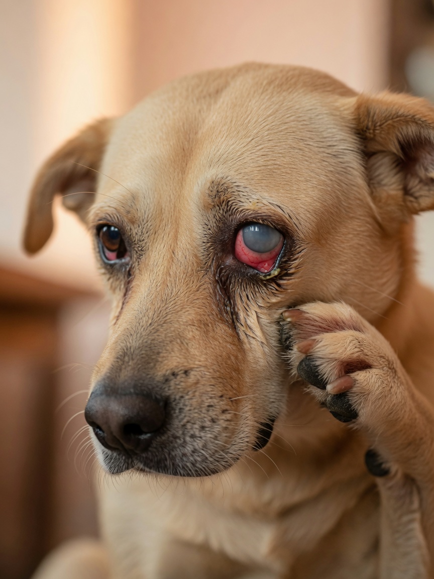
The clinical signs of acute glaucoma in dogs include a red or bloodshot eye, cloudiness or a blue-grey haze over the cornea, a visibly enlarged or bulging eyeball (buphthalmos in chronic cases), squinting or sensitivity to light, apparent pain including face rubbing, reduced appetite, and lethargy. Dogs in acute glaucoma are often genuinely distressed, though some stoic individuals hide their discomfort well. Any eye that is red, cloudy, and appears painful warrants emergency veterinary assessment the same day — do not wait to see if it resolves overnight.
Diagnosis and Pressure Measurement
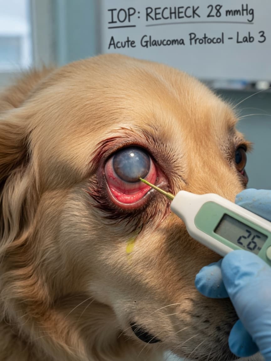
Intraocular pressure is measured using a tonometer — a small probe touched briefly to the surface of the anaesthetised cornea. This is a quick, minimally uncomfortable procedure. Pressures above 25 to 30 mmHg are suspicious; pressures above 40 to 50 mmHg are consistent with acute glaucoma and represent a true emergency. The vet will also examine the drainage angle using a special contact lens (gonioscopy) to determine whether the condition is primary or secondary, and will assess the degree of optic nerve and retinal damage.
Treatment Approaches
Emergency Management
The immediate priority is reducing intraocular pressure as rapidly as possible to limit further damage to the optic nerve and retina. This typically involves intravenous mannitol to rapidly draw fluid from the eye, combined with topical and oral medications to reduce aqueous production. How much vision can be salvaged depends almost entirely on how elevated the pressure was and for how long before treatment began.
Long-Term Medical Management
Once pressure is stabilised, long-term management typically involves a combination of topical carbonic anhydrase inhibitors and prostaglandin analogues to reduce aqueous production and improve drainage. These are administered as eye drops, often twice daily. Regular tonometry rechecks are essential to confirm pressure remains controlled.
Surgical Options
Medical management alone is frequently insufficient for long-term pressure control. Surgical options include laser cyclophotocoagulation to reduce aqueous production, gonioimplant (drainage shunt) insertion, and in eyes where vision has already been lost, procedures to relieve pain without preserving sight — such as chemical ablation of the ciliary body or enucleation. A referral to a veterinary ophthalmologist is strongly advisable for all but the most straightforward cases.
Prophylactic Treatment of the Fellow Eye
In dogs diagnosed with primary glaucoma, veterinary ophthalmologists commonly recommend starting prophylactic eye drops in the unaffected eye to delay or prevent disease onset. This is a well-supported approach given the high risk of bilateral disease.
Key Takeaways
- Acute glaucoma in dogs is an emergency: vision can be lost within 24 to 72 hours of pressure rising
- A red, cloudy, painful eye requires same-day veterinary assessment — never wait overnight
- Several breeds have inherited predisposition to primary glaucoma; the second eye is also at significant risk
- Tonometry is the definitive diagnostic tool and should be performed in any dog with a suspect eye
- Long-term management requires ongoing medication and regular rechecks; referral to a specialist is advisable
- Prophylactic treatment of the unaffected eye is recommended in confirmed primary glaucoma cases