
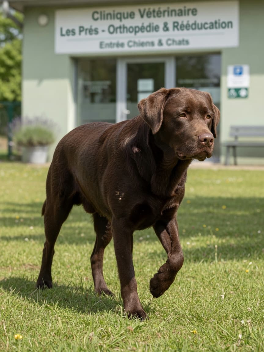
What Is Osteochondrosis?
Osteochondrosis — often abbreviated to OC, or OCD when a cartilage flap has separated — is a disturbance in the normal process by which cartilage is converted into bone during skeletal development. In a healthy growing puppy, cartilage cells in the joints follow a precise sequence: they proliferate, mature, receive nutrients through blood vessels, and eventually calcify to form bone. In osteochondrosis, this sequence is disrupted. A localised area of cartilage fails to receive adequate blood supply, the cells die, and a thickened, abnormal cartilage lesion develops on the surface of the joint.
When the degenerated cartilage cracks and lifts away from the underlying bone, the condition is called osteochondrosis dissecans (OCD). The detached flap — sometimes referred to as a joint mouse — floats within the joint space, causing inflammation, pain, and cartilage erosion. Even before a flap forms, the weakened cartilage causes discomfort and initiates arthritic changes that can persist throughout the dog's life.
Why Does It Happen?
Osteochondrosis is understood to arise from a combination of genetic susceptibility and environmental factors, particularly those associated with rapid growth. The two most significant contributors are:
- Genetics — certain bloodlines within large and giant breeds have a much higher incidence of OCD, and the condition clusters in families
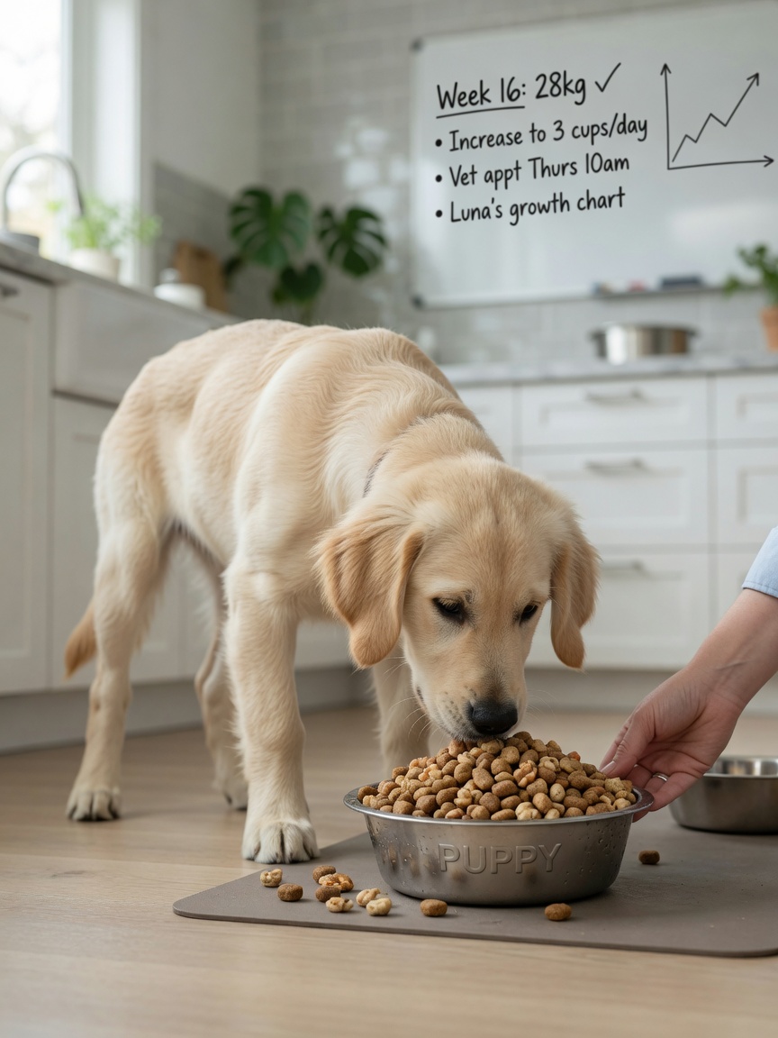
- Rapid growth — puppies fed high-calorie diets that promote faster-than-normal growth rates are at increased risk, as the cartilage may not mature quickly enough to keep pace with skeletal expansion
Excessive dietary calcium and phosphorus supplementation in puppies fed large breed food has historically been implicated as well, though modern large breed puppy diets are formulated to reduce this risk. Trauma to developing cartilage may also play a role in triggering or accelerating lesion formation in susceptible individuals.
Breeds Most Commonly Affected
Osteochondrosis is overwhelmingly a disease of large and giant breed dogs. Breeds with the highest reported incidence include:
- Labrador Retrievers
- Golden Retrievers
- Great Danes
- Bernese Mountain Dogs
- Rottweilers
- Irish Wolfhounds
- Border Collies
Males are more frequently affected than females, which may relate to their faster growth rate or hormonal influences on cartilage maturation. Most dogs develop clinical signs between five and eight months of age, coinciding with the period of most rapid skeletal growth.
Sites of Osteochondrosis
While osteochondrosis can affect any joint where cartilage undergoes the endochondral ossification process, certain locations are far more common than others.
The Shoulder — Most Common Site
The caudal (rear) aspect of the humeral head — the ball of the shoulder joint — is by far the most frequently affected location. The caudal humeral head is a site of significant mechanical stress during locomotion, and lesions here tend to produce the most clinically obvious lameness. Dogs with shoulder OCD typically present with a consistent forelimb lameness that is worse after exercise, pain on extension and flexion of the shoulder, and muscle wastage in the affected limb over time.
The Elbow
OCD of the elbow, particularly affecting the medial humeral condyle, falls under the broader umbrella of elbow dysplasia. It is a significant contributor to forelimb lameness in young large breeds and often occurs alongside other components of elbow dysplasia such as fragmented coronoid process.
The Hock (Tarsus)
Tarsal OCD most commonly affects the medial trochlear ridge of the talus. It tends to present slightly later than shoulder or elbow OCD and can cause a characteristic hind limb lameness in young dogs. Rottweilers are particularly predisposed to tarsal OCD.
The Stifle (Knee)
Stifle OCD is less common than shoulder or elbow involvement but does occur. Lesions are typically found on the femoral condyles and can be difficult to distinguish from other causes of stifle pathology without advanced imaging.
Diagnosing Osteochondrosis
Diagnosis begins with a thorough orthopaedic examination. Vets will assess gait, palpate the affected joint for pain and effusion, and test the range of motion. Radiographs — X-rays — are the first-line imaging modality and will often reveal a flattening or concavity on the articular surface of the affected bone, along with joint effusion and sometimes a visible flap fragment.
In early cases or where X-ray findings are subtle, advanced imaging with CT (computed tomography) or MRI provides much greater detail of the cartilage and bone. CT in particular is widely used in specialist centres to characterise the size and location of lesions before planning surgery.
Treatment: Why Arthroscopy Is Preferred
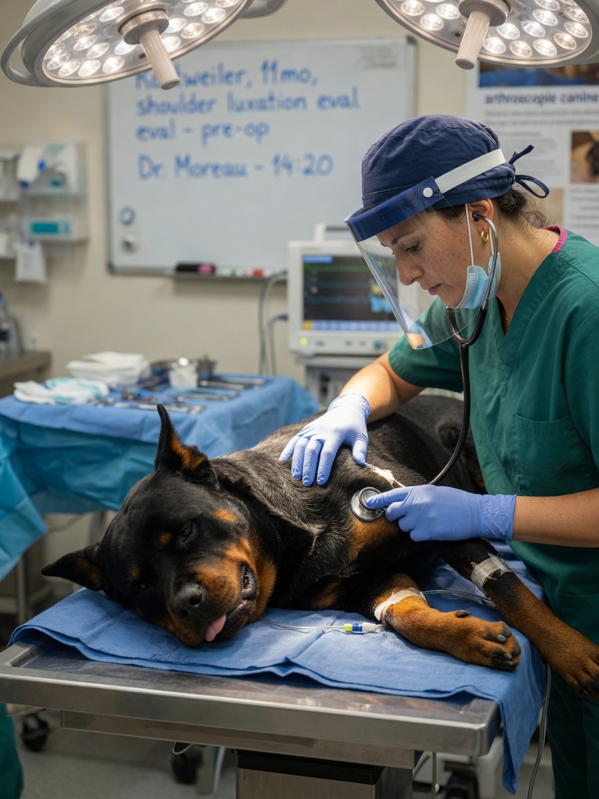
For the vast majority of dogs with OCD showing clinical signs, surgical treatment is recommended. The goal is to remove the loose cartilage flap and any loose fragments, debride the underlying subchondral bone to encourage fibrocartilage healing, and assess the overall joint for associated pathology.
Arthroscopic surgery has become the preferred technique at referral centres across the UK. Using a small camera and operating instruments introduced through tiny portals in the joint capsule, the surgeon can visualise the entire joint, remove fragments precisely, and manage the lesion with minimal disruption to surrounding tissues. Recovery times are shorter, post-operative pain is reduced, and the surgeon has excellent visibility of the joint compared to open approaches.
Open arthrotomy — where the joint is opened through a larger incision — remains an option and may still be preferred in some cases, particularly for hock lesions where joint access via arthroscope can be more challenging.
Prognosis and Long-Term Outlook
The prognosis for dogs with shoulder OCD treated surgically is generally very good, particularly when surgery is performed before significant secondary arthritis has developed. Most dogs return to a good level of function, though some degree of joint change is inevitable and may cause intermittent discomfort in later life.
For OCD of the elbow, hock, and stifle, outcomes are more variable and depend on the severity of secondary changes at the time of treatment. Early intervention consistently yields better outcomes than delayed treatment. Post-operative rehabilitation, including controlled lead exercise and physiotherapy, is important in all cases to ensure the best possible recovery and to rebuild muscle mass lost through the period of lameness.
Feeding a large breed specific puppy food — which is formulated to support steady, controlled growth rather than rapid weight gain — is one of the most practical preventive measures owners of at-risk breeds can take.