
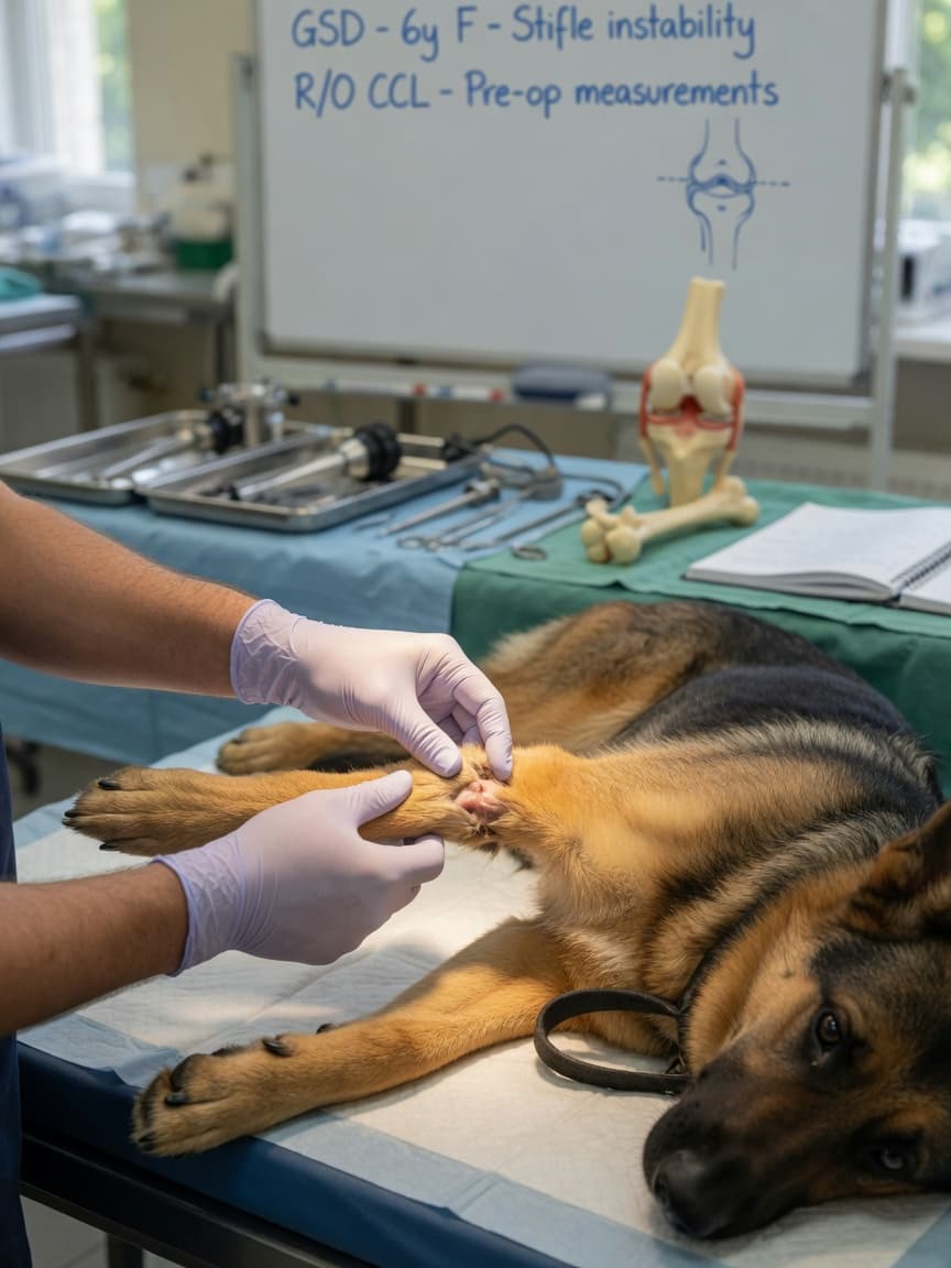
The Diagnosis That Changes Everything
Cranial cruciate ligament rupture is the single most common orthopaedic injury in dogs. It accounts for more referrals to veterinary orthopaedic specialists than any other condition, and the associated costs to dog owners in the UK alone run into hundreds of millions of pounds annually. When a dog suddenly goes lame on a hindlimb — or gradually worsens over weeks — cruciate disease is high on the differential list. Understanding the surgical options available is the first step in making an informed decision for your dog.
What the Cruciate Ligament Does and Why It Fails
The cranial cruciate ligament (CrCL) sits inside the stifle joint — the canine equivalent of the human knee — and prevents the tibia from sliding forward relative to the femur during weight-bearing. Unlike in humans, where cruciate rupture is typically a sudden trauma, dogs most commonly suffer a degenerative process: the ligament weakens progressively due to a combination of genetic predisposition, conformation, body weight, and immune-mediated factors, until partial or complete rupture occurs.
Once the CrCL is compromised, the stifle becomes unstable. This instability causes pain, inflammation, rapid onset of osteoarthritis, and in many cases concurrent meniscal damage — the cartilage pads within the joint are frequently torn as a secondary injury.
Why Surgery Is Recommended
Unlike some ligament injuries in other species, canine CrCL rupture does not reliably heal with rest alone. In small dogs under fifteen kilograms, conservative management with strict rest and physiotherapy can produce acceptable outcomes in some cases. In medium, large, and giant breeds, surgery is considered the standard of care. Without surgical stabilisation, ongoing instability accelerates joint degeneration and the dog remains in pain and functionally limited.
Three main surgical techniques are in widespread use: the tibial plateau levelling osteotomy (TPLO), the tibial tuberosity advancement (TTA), and the lateral suture technique (also called extracapsular repair or fabella-tibial suture). Each works differently, and each has a place depending on patient factors.
TPLO: Tibial Plateau Levelling Osteotomy
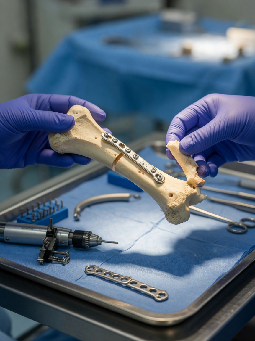
TPLO is currently the most widely performed cruciate repair technique at specialist referral centres. Rather than replacing the torn ligament, it changes the geometry of the tibia so that the ligament is no longer needed for stability during normal gait. The surgeon cuts a circular arc through the top of the tibia (the tibial plateau), rotates the bone segment to reduce the slope of the plateau, and secures it with a bone plate and screws. Once the new angle is established, the quadriceps muscles provide active stabilisation without relying on a ligament.
TPLO produces consistently excellent outcomes in dogs of all sizes, including large and giant breeds. Return to full activity typically occurs at sixteen weeks, and long-term function is generally very good. It requires specialist surgical training and equipment, and cost reflects this.
TTA: Tibial Tuberosity Advancement
TTA works on a related biomechanical principle: by advancing the tibial tuberosity (the bony prominence at the front of the tibia where the patellar tendon attaches), the surgeon aligns the patellar tendon perpendicular to the tibial plateau, neutralising the forward sliding force on the tibia. A cage, fork, and plate system stabilises the advancement while bone heals.
TTA is broadly comparable to TPLO in outcomes for many patient populations, and some surgeons favour it for dogs with certain tibial conformations. Recovery time is similar, and the procedure also requires specialist training. Some studies suggest TTA may carry a slightly higher rate of certain post-operative complications compared to TPLO, though evidence continues to evolve and the difference is not dramatic in experienced hands.
Lateral Suture: Extracapsular Repair
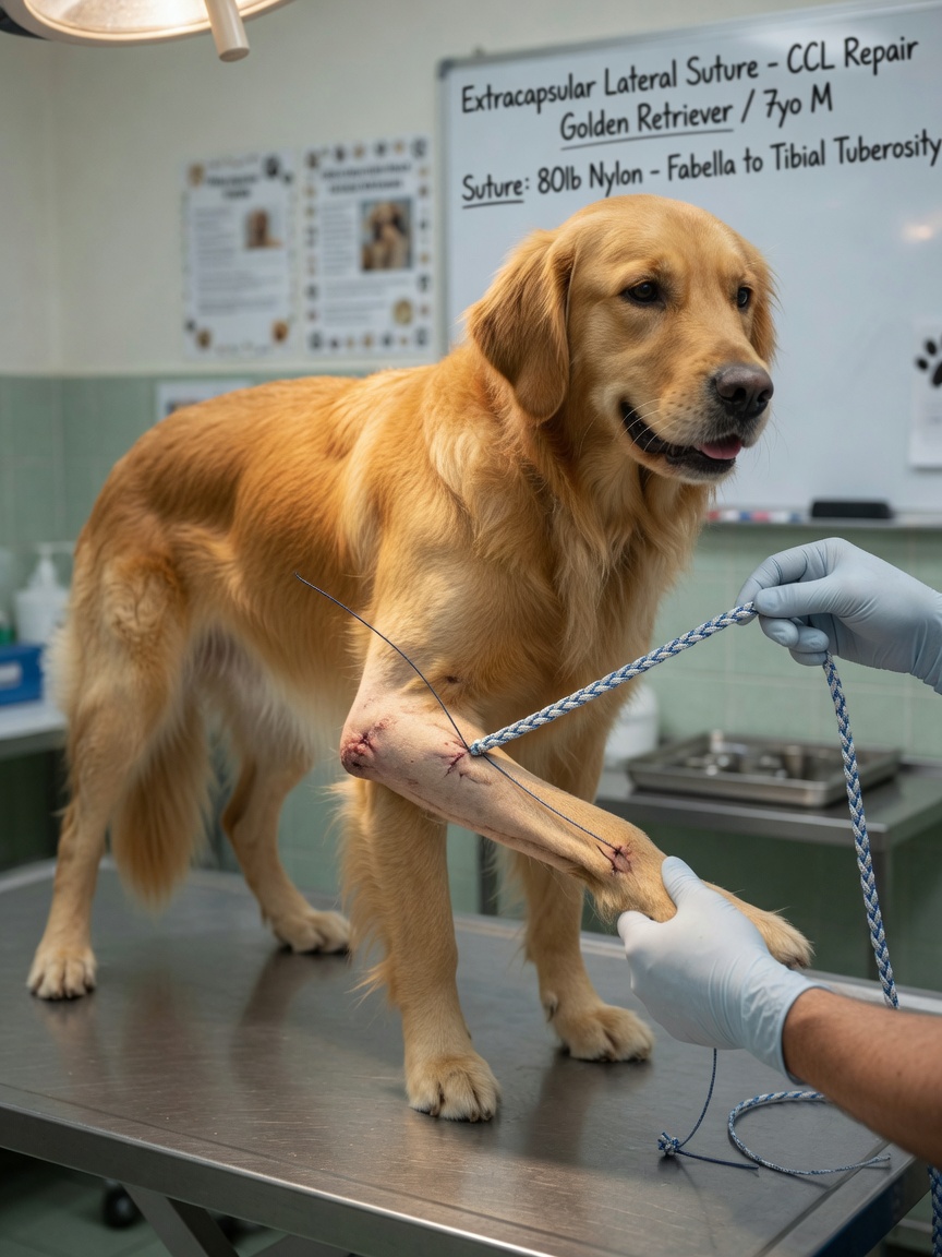
The lateral suture technique takes an entirely different approach. Rather than modifying bone geometry, the surgeon places a heavy nylon suture outside the joint capsule, running from the lateral fabella (a small sesamoid bone behind the femur) to a point on the tibial crest. This suture mimics the action of the CrCL, restraining forward tibial movement while scar tissue forms and provides longer-term stability.
The suture itself is expected to eventually break or stretch — the goal is to maintain stability long enough for a fibrous tissue response to develop. This technique is technically more accessible, less expensive, and available at many general practices without specialist referral.
For dogs under fifteen to twenty kilograms, lateral suture outcomes are generally comparable to TPLO and TTA. In larger, more active, or heavier dogs, re-rupture or suture failure rates are higher, and bone-based techniques tend to produce more reliable long-term stability.
Choosing Between the Three
There is no universally correct answer — the best procedure depends on your dog's size, conformation, activity level, tibial plateau angle, and your access to specialist surgical services.
- Large and giant breeds: TPLO or TTA are strongly preferred due to the forces involved.
- Small dogs under fifteen kilograms: lateral suture is a reasonable, cost-effective option with good outcomes.
- Dogs with steep tibial plateau angles: TPLO is particularly well-suited.
- Budget constraints with a small dog: lateral suture at an experienced general practitioner is a legitimate choice.
- High-performance or working dogs: TPLO tends to be the preferred choice for return to demanding physical activity.
Meniscal damage should always be assessed at the time of surgery; if a torn meniscus is found, it is treated concurrently. Failure to address meniscal injury is a recognised cause of ongoing post-operative pain.
What Owners Can Expect After Surgery
Regardless of technique, recovery involves a period of strict rest, followed by gradual reintroduction of controlled exercise and physiotherapy. Hydrotherapy is particularly valuable in the recovery period. Most dogs return to full activity by four to five months. The operated limb will develop some degree of osteoarthritis over time, but surgical stabilisation significantly slows its progression compared to an unstabilised stifle.
Always consult a veterinary orthopaedic specialist to determine which procedure is most appropriate for your dog's specific anatomy and lifestyle requirements.