
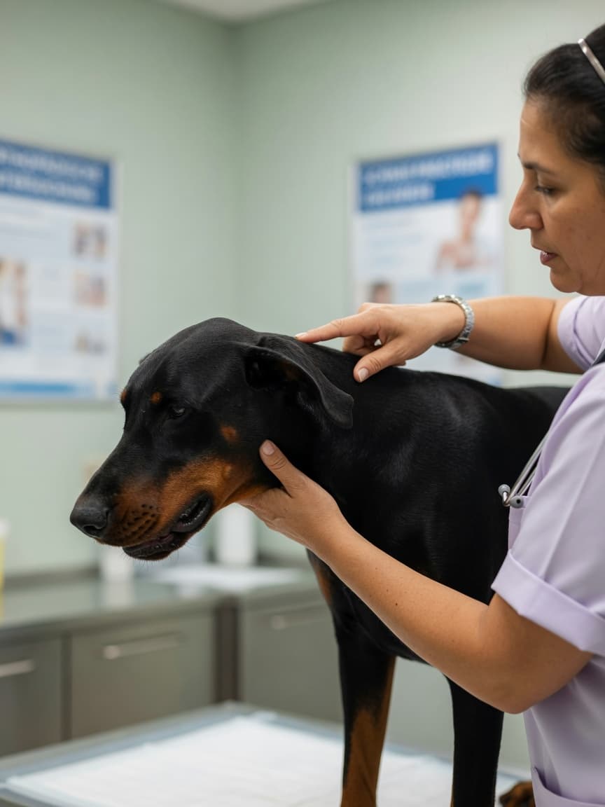
Wat is Wobbler Syndroom?
Wobbler syndroom is de gangbare naam voor cervicale spondylomyelopathie (CSM) — een aandoening waarbij afwijkingen van de cervicale (nek)wervelkolom het ruggenmerg of zenuwwortels in het nekgebied comprimeren. De compressie verstoort de overdracht van zenuwsignalen tussen de hersenen en de ledematen, wat resulteert in de karakteristieke wankele, onvaste gang die de aandoening zijn naam geeft.
CSM is geen enkele, uniforme ziekte. Het omvat een reeks structurele afwijkingen, waaronder hernia van de tussenwervelschijf, instabiliteit van de wervelkolom, benige vormafwijking van de wervels en hypertrofie (verdikking) van ligamenten in het ruggenmergkanaal. Het eindresultaat is in elk geval hetzelfde: vernauwing van het ruggenmergkanaal en compressie van het delicate weefsel daarbinnen.
Raspredisposities
Wobbler syndroom vertoont een duidelijke voorkeur voor grote en reuzenrassen, waarbij twee in het bijzonder opvallen:
Dobermann
De Dobermann is het ras dat het meest wordt getroffen door wobbler syndroom, en vormt in veel onderzoeken de meerderheid van de gevallen. Bij dit ras betreft de aandoening meestal schijf-gerelateerde compressie, meestal op de C5-C6 of C6-C7 aansluiting. Het ontstaan is meestal op middelbare leeftijd, tussen vijf en negen jaar, hoewel jongere honden kunnen worden getroffen. De aandoening bij Dobermanns is meestal chronisch en progressief.
Great Dane
Great Danes ontwikkelen een vorm van CSM die verschilt van het Dobermann-type. Het betreft vaker benige vormafwijking van de wervellichamen zelf — soms het "wobbler" of "schijf" type versus het "benige" type genoemd — en treft meestal jongere honden, vaak tussen een en drie jaar oud. Andere reuzenrassen zoals de Mastiff, Sint-Bernard en Ierse Wolfhond zijn ook vertegenwoordigd.
Klinische Verschijnselen
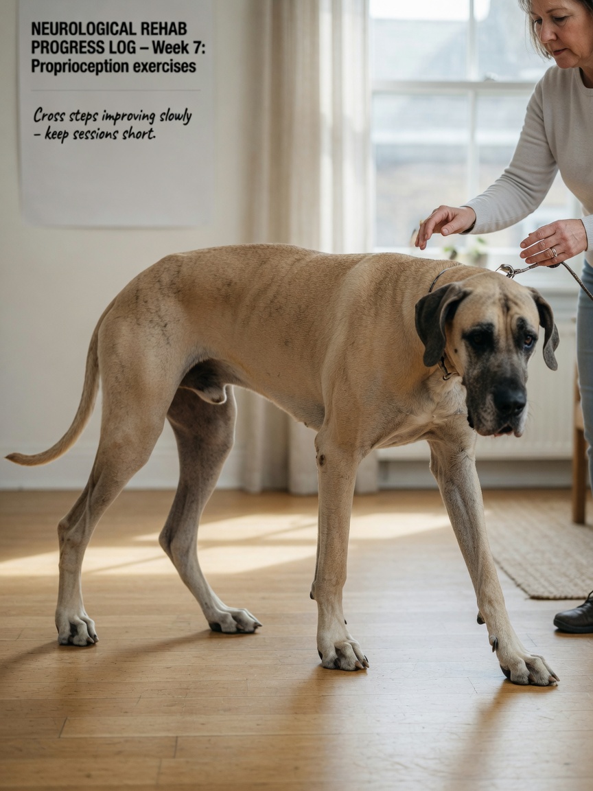
De presentatie van wobbler syndroom weerspiegelt de locatie en ernst van de ruggenmergcompressie.
Proprioceptieve Tekorten en Wankele Gang
Het meest karakteristieke teken is een abnormale gang in de achterpoten. Aangetaste honden lijken te struikelen of te zwaaien, zetten hun poten ongelijk neer en kruisen hun achterpoten soms terwijl ze lopen. Dit gebeurt omdat proprioceptie — het sensorische bewustzijn van waar de ledematen zich in de ruimte bevinden — wordt belemmerd door de compressie. Door de achterkant van de poot van de hond op de grond te plaatsen en te kijken hoe snel dit wordt gecorrigeerd (de proprioceptieve plaatsingstest), worden vaak duidelijke tekorten opgeleverd. De voorpoten kunnen ook worden getroffen, maar ataxie van de achterpoten is meestal meer uitgesproken omdat de relevante ruggenmergbanen selectief kwetsbaar zijn.
Neckpijn
Veel honden met wobbler syndroom vertonen tekenen van neckpijn. Ze kunnen terughoudend zijn om hun hoofd naar beneden te laten zakken om te eten of drinken, verzetten zich tegen buiging of extensie van de nek, of schreeuwen wanneer de nek wordt palpeerd. Sommige honden nemen een karakteristieke, stijf gehouden kophouding aan. Neckpijn kan onderbroken zijn en is niet altijd duidelijk bij een enkel onderzoek.
Betrokkenheid van Voorpoten
Wanneer de compressie ernstig is of meerdere plaatsen betreft, kunnen de voorpoten zwakte, stijfheid of verkorte stap vertonen — soms omschreven als een "hakkelende" voorpotgang. In de meest ernstige gevallen kunnen honden niet in staat zijn om te lopen of kunnen zich presenteren als tetraparetisch (zwakte in alle vier ledematen).
Diagnose
Gewone röntgenafbeeldingen van de cervicale wervelkolom kunnen de diagnose suggereren door abnormale afstand tussen wervels, benige veranderingen of verminderde schijfhoogte aan het licht te brengen. MRI is echter de gouden standaard voor het bepalen van de precieze locatie, aard en mate van ruggenmergcompressie. Het biedt het detail dat nodig is voor chirurgische planning en helpt bepalen of één of meerdere plaatsen zijn betrokken — een belangrijk onderscheid dat de behandelingskeuze beïnvloedt.
Myelografie (inspuiting van contrastmiddel in het ruggenmergvocht om het ruggenmergkanaal onder röntgenstraling uit te lichten) was de historische onderzoeksmethode van keuze voordat MRI algemeen beschikbaar werd en wordt nog steeds in sommige praktijken gebruikt. Dynamische myelografie — het afbeelden van de nek in zowel buiging als extensie — kan compressie openbaren die slechts in bepaalde posities aanwezig is.
Behandelingsopties
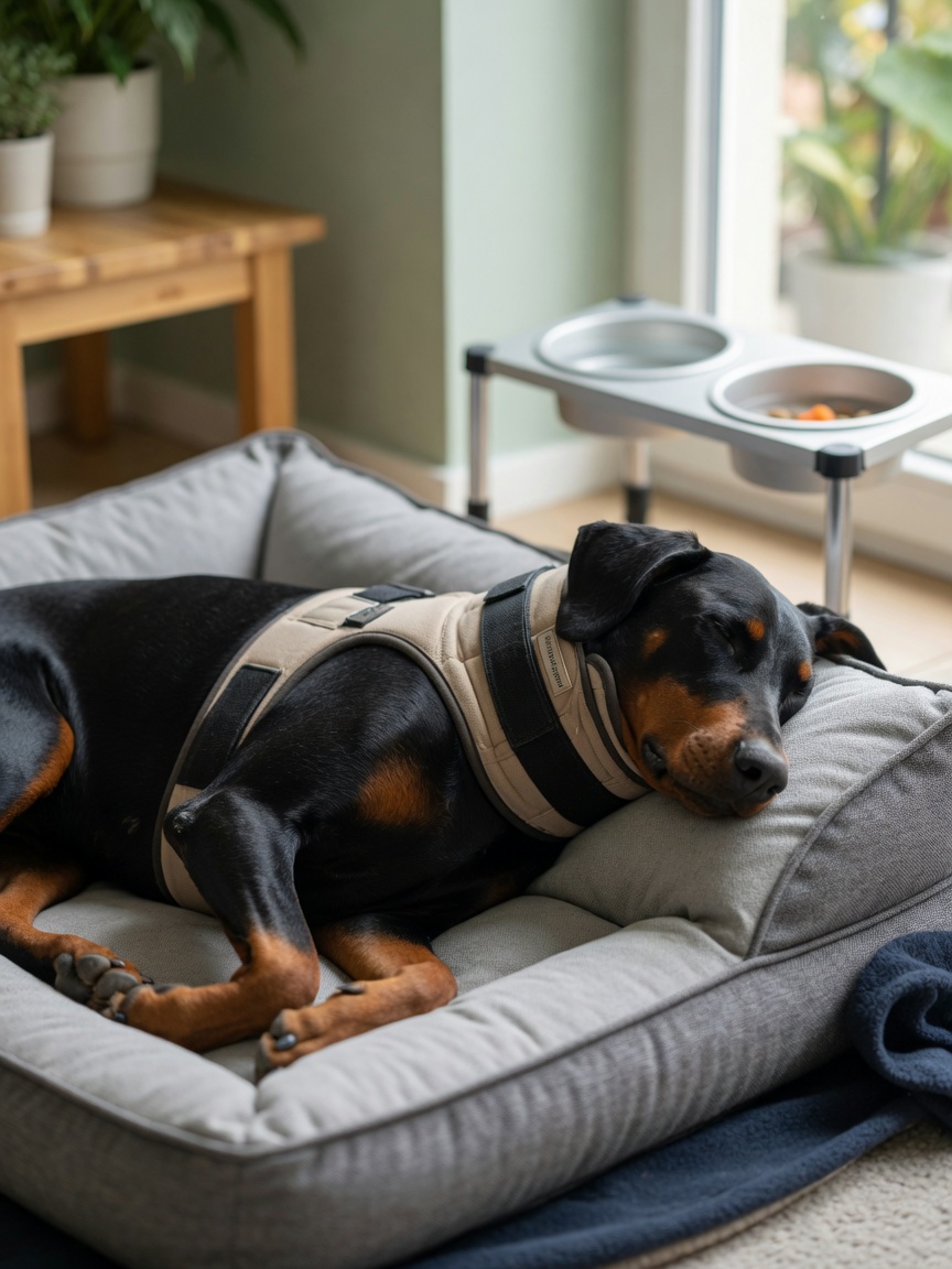
Conservatief Beheer
Conservatief beheer is gericht op het verminderen van ontstekingen en het beperken van beweging zodat het ruggenmerg kan herstellen van de compressie. Het omvat doorgaans een periode van streng rust, ontstekingsremmende medicatie en pijnverlichting. Een neksteun kan worden gebruikt om cervicale beweging te beperken en stress op de gecomprimeerde segmenten te verminderen.
Conservatief beheer is het meest geschikt voor honden met milde tot matige verschijnselen, honden die te groot risico vormen voor chirurgie vanwege andere gezondheidsomstandigheden, of eigenaren die geen chirurgische interventie kunnen volgen. Ongeveer de helft van de honden die conservatief worden behandeld, vertonen betekenisvolle verbetering, hoewel recidieven en progressie veel voorkomen.
Chirurgische Opties
Chirurgie is gericht op decompressie van het ruggenmerg en, waar van toepassing, stabilisatie van de aangetaste wervelstukken. Verschillende chirurgische benadering zijn beschreven, waarbij de keuze afhangt van de aard van de compressie, het aantal betrokken plaatsen en de individuele anatomie van de hond.
De ventrale sleuvenoperatie is een van de meest uitgevoerde technieken voor schijf-gerelateerde compressie. De chirurg nadert de cervicale wervelkolom van onder de nek, verwijdert een klein venster van bot en schijfmateriaal en decomprimieert het ruggenmerg rechtstreeks. Het is technisch veeleisend maar goed gevestigd, met goede resultaten gerapporteerd in veel series.
Distraction-fusietechnieken worden gebruikt om aangetaste segmenten te stabiliseren terwijl tegelijkertijd de compressie wordt opgeheven. Implantaten worden geplaatst om de wervels uit elkaar te spreiden en de correctie in stand te houden terwijl benige fusie optreedt in de daaropvolgende weken tot maanden. Deze procedures hebben meer steun gekregen voor ziekten met meerdere plaatsen en voor gevallen waarbij eenvoudige decompressie onvoldoende zal zijn.
Prognose
De prognose voor wobbler syndroom hangt sterk af van de ernst van verschijnselen bij presentatie, het aantal betrokken plaatsen en de gevolgde behandeling. Honden met milde tot matige verschijnselen die succesvol decompressieve chirurgie ondergaan, doen het doorgaans goed, en een aanzienlijk deel vertoont duidelijke verbetering. Honden die niet kunnen lopen op het moment van chirurgie hebben een meer voorzichtige outlook. Recidief op dezelfde of aangrenzende plaatsen is een erkende complicatie, vooral bij honden die over lange termijn worden behandeld.
Regelmatige fysiotherapie en watergymnastiek zijn waardevolle aanvullingen op zowel chirurgische als conservatieve behandeling, helpen spiermatstelling en proprioceptieve functie tijdens behandeling en herstel in stand te houden.