
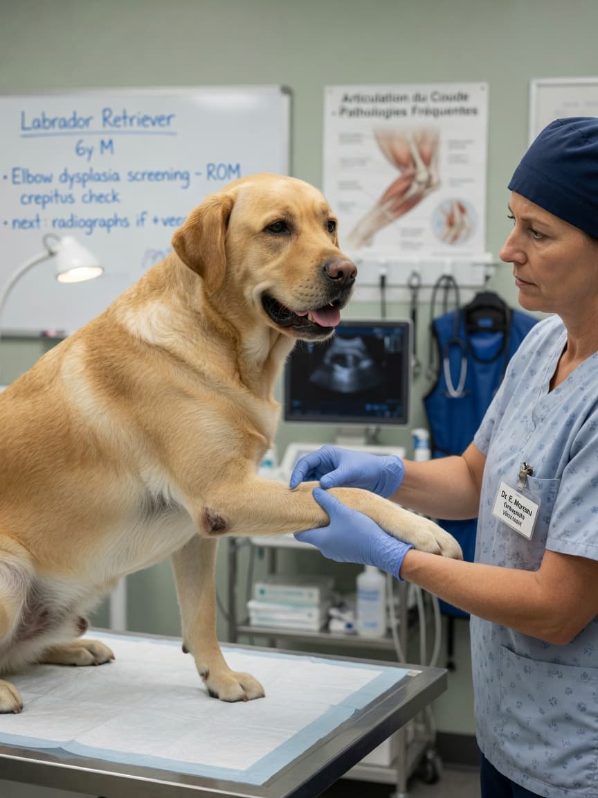
The Elbow Condition That Is Often Missed Until Damage Is Done
Elbow dysplasia is the most common cause of forelimb lameness in large breed dogs, yet it is frequently underdiagnosed in its early stages. Studies suggest it affects up to forty percent of Labrador Retrievers, with German Shepherds, Golden Retrievers, Rottweilers, and Bernese Mountain Dogs also significantly represented. Unlike hip dysplasia, elbow dysplasia encompasses several distinct pathological processes within the same joint — and distinguishing between them directly influences treatment decisions.
Understanding Elbow Dysplasia: It Is Not One Disease
The term elbow dysplasia describes a group of developmental conditions affecting the elbow joint during growth. The four recognised components are fragmented medial coronoid process, osteochondrosis dissecans of the medial humeral condyle, ununited anconeal process, and medial compartment disease. These conditions often occur together, and all result in abnormal contact mechanics within the joint, progressive cartilage damage, and osteoarthritis.
A fragmented medial coronoid process is the most common presentation. A small fragment of bone within the joint causes inflammation, synovitis, and cartilage erosion on the opposing humeral surface. Osteochondrosis dissecans involves a flap of damaged cartilage that separates from the underlying bone, releasing debris into the joint. An ununited anconeal process occurs when a secondary ossification centre fails to fuse, leaving an unstable bony fragment at the back of the elbow.
Recognising the Clinical Signs
Most affected dogs begin showing signs between four and ten months of age, though some cases are not identified until adulthood when secondary arthritis becomes symptomatic. The classic presentation is a forelimb lameness that worsens after exercise and improves with rest, only to return with activity. Dogs may hold the affected limb slightly away from the body, show reduced range of motion on examination, or flinch when the elbow is flexed or extended fully.
Bilateral involvement is common — roughly thirty to fifty percent of affected dogs have disease in both elbows simultaneously, which can make lameness appear less pronounced or even symmetrically distributed, delaying recognition. Any large breed puppy showing subtle or intermittent forelimb stiffness should have elbows evaluated, not just hips.
Diagnosis: Beyond Physical Examination
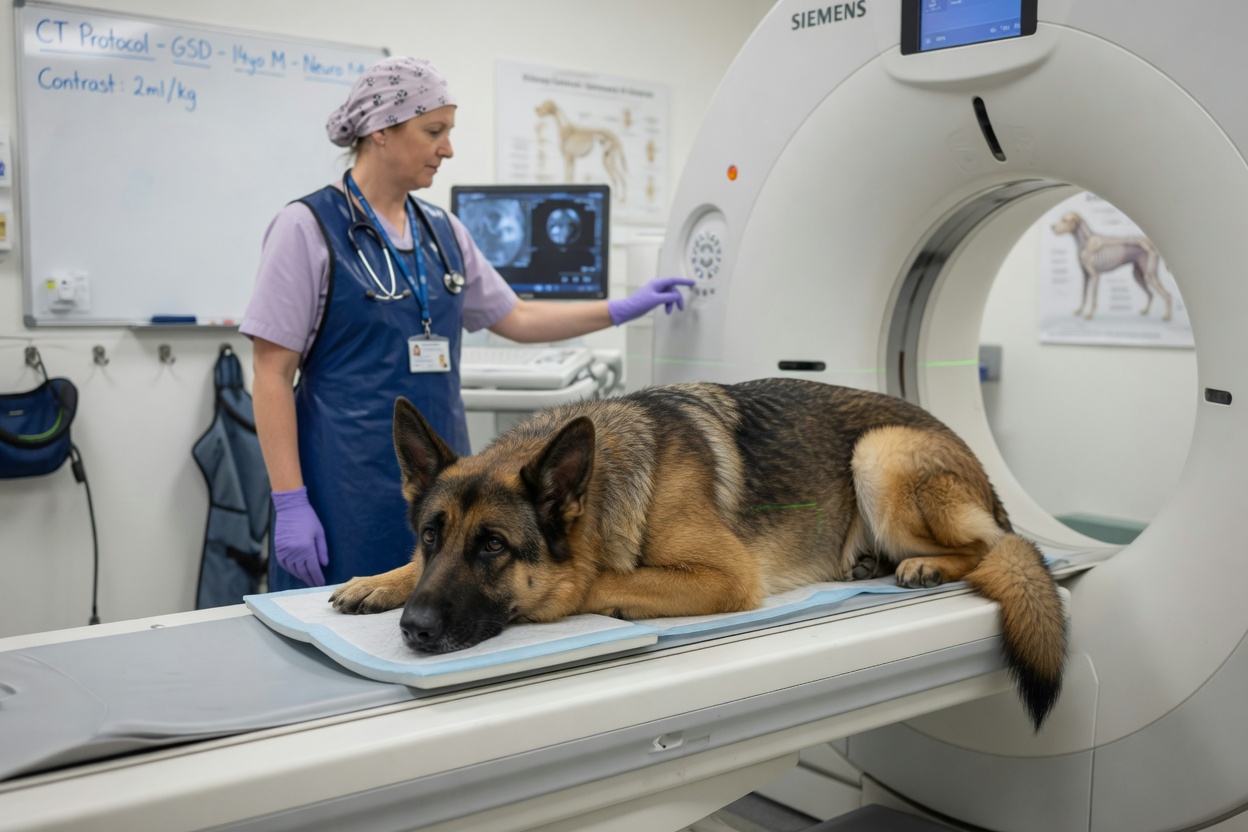
Standard radiography can identify ununited anconeal process and more advanced arthritic changes, but it frequently underestimates the extent of fragmented coronoid process disease and cartilage damage. Computed tomography has become the diagnostic standard for elbow dysplasia, providing three-dimensional detail of bony structures that plain radiographs cannot capture. Many specialist centres now recommend CT of both elbows at the time of initial diagnosis to determine whether bilateral disease is present and to plan surgical approach accurately.
Arthroscopy — minimally invasive joint camera examination — simultaneously serves as both a diagnostic and a therapeutic tool. It allows direct visualisation of cartilage surfaces, identification and removal of fragments, and assessment of medial compartment disease that influences long-term prognosis.
Surgical Management
Arthroscopic fragment removal
For fragmented medial coronoid process and osteochondrosis dissecans flaps, arthroscopic removal of loose fragments and debridement of damaged cartilage is the most widely performed procedure. Evidence supports better outcomes with arthroscopy compared to open surgery due to reduced post-operative morbidity and superior visualisation. Early intervention — before significant cartilage erosion has occurred — consistently produces better functional results.
Proximal abducting ulnar osteotomy
Where significant medial compartment disease is present — meaning erosion of both the medial coronoid cartilage and the opposing humeral surface — fragment removal alone has limited benefit. Procedures designed to unload the medial compartment, such as proximal abducting ulnar osteotomy, aim to redistribute joint contact forces and slow cartilage degeneration. These are more complex interventions typically performed by veterinary orthopaedic specialists.
Ununited anconeal process repair
Surgical options for an ununited anconeal process include lag screw fixation to encourage fusion, or fragment removal combined with a dynamic proximal ulnar osteotomy to address underlying incongruence. Which approach is appropriate depends on the dog's age and the degree of existing secondary changes.
Conservative Management
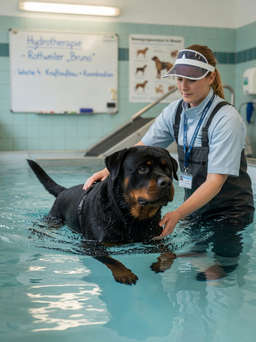
Not all dogs are surgical candidates, and owners sometimes choose conservative management either by preference or because advanced arthritis means surgery offers limited additional benefit. Conservative management centres on multimodal pain control, weight management, controlled physiotherapy, and targeted exercise modification.
Non-steroidal anti-inflammatory drugs are the cornerstone of medical management when prescribed and monitored by a veterinarian. Joint supplements containing omega-3 fatty acids have supporting evidence for modest anti-inflammatory effects in canine osteoarthritis. Hydrotherapy and land-based physiotherapy help maintain muscle mass and joint range of motion. Keeping the dog at an ideal body weight is arguably the single most impactful conservative intervention, as even small reductions in body mass significantly reduce joint loading forces.
It is important to understand that conservative management controls symptoms and slows progression — it does not address the underlying structural problem. Dogs managed conservatively require long-term veterinary monitoring and honest reassessment if quality of life deteriorates.
Long-Term Outlook and What Owners Should Expect
Elbow dysplasia is a lifelong condition. Even after successful surgery, the underlying predisposition to osteoarthritis remains, and most affected dogs will require some degree of ongoing management as they age. Early surgical intervention in young dogs with minimal secondary changes offers the best functional outcomes, with many dogs returning to comfortable activity levels.
- Consult a veterinary orthopaedic specialist if elbow dysplasia is suspected — general practice radiographs often underestimate severity
- Request CT imaging to assess both elbows and guide surgical planning
- Discuss arthroscopy as the preferred surgical approach for fragment removal
- Begin physiotherapy and hydrotherapy as part of post-operative rehabilitation
- Maintain lean body condition throughout the dog's life
- Schedule regular veterinary reviews to monitor osteoarthritis progression and adjust pain management accordingly