
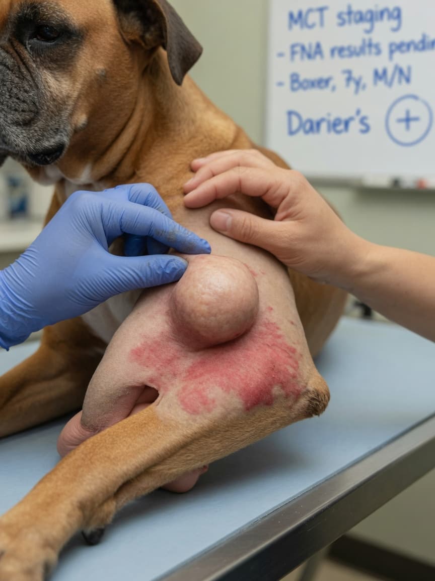
What Are Mast Cell Tumours?
Mast cell tumours (MCTs) arise from mast cells, which are immune cells found throughout the body's connective tissue and skin. Mast cells play a role in allergic responses and wound healing, and they contain granules packed with biologically active substances including histamine, heparin, and various inflammatory mediators. When these cells become cancerous, they form tumours that can release these substances — a process called degranulation — which is central to many of the clinical challenges associated with treating MCTs.
Mast cell tumours are the single most common skin tumour diagnosed in dogs, accounting for between 16 and 21 per cent of all canine skin tumours. They can occur anywhere on the body but are most frequently found on the trunk, limbs, and perineal region.
Breeds Most at Risk
Certain breeds show a markedly higher predisposition to mast cell tumours, suggesting a genetic component to their development. Brachycephalic breeds are particularly overrepresented:
- Boxers — one of the highest-risk breeds, though their tumours tend to be lower grade and carry a better prognosis
- English Bulldogs
- Boston Terriers
- French Bulldogs
Retriever breeds also carry elevated risk:
- Labrador Retrievers
- Golden Retrievers
Flat-Coated Retrievers are notable in that they tend to develop higher-grade, more aggressive MCTs than other retriever breeds. MCTs can develop in any breed and in mixed-breed dogs, so owners should not discount a suspicious lump simply because their dog is not on a predisposed breed list.
Clinical Appearance and Darier's Sign
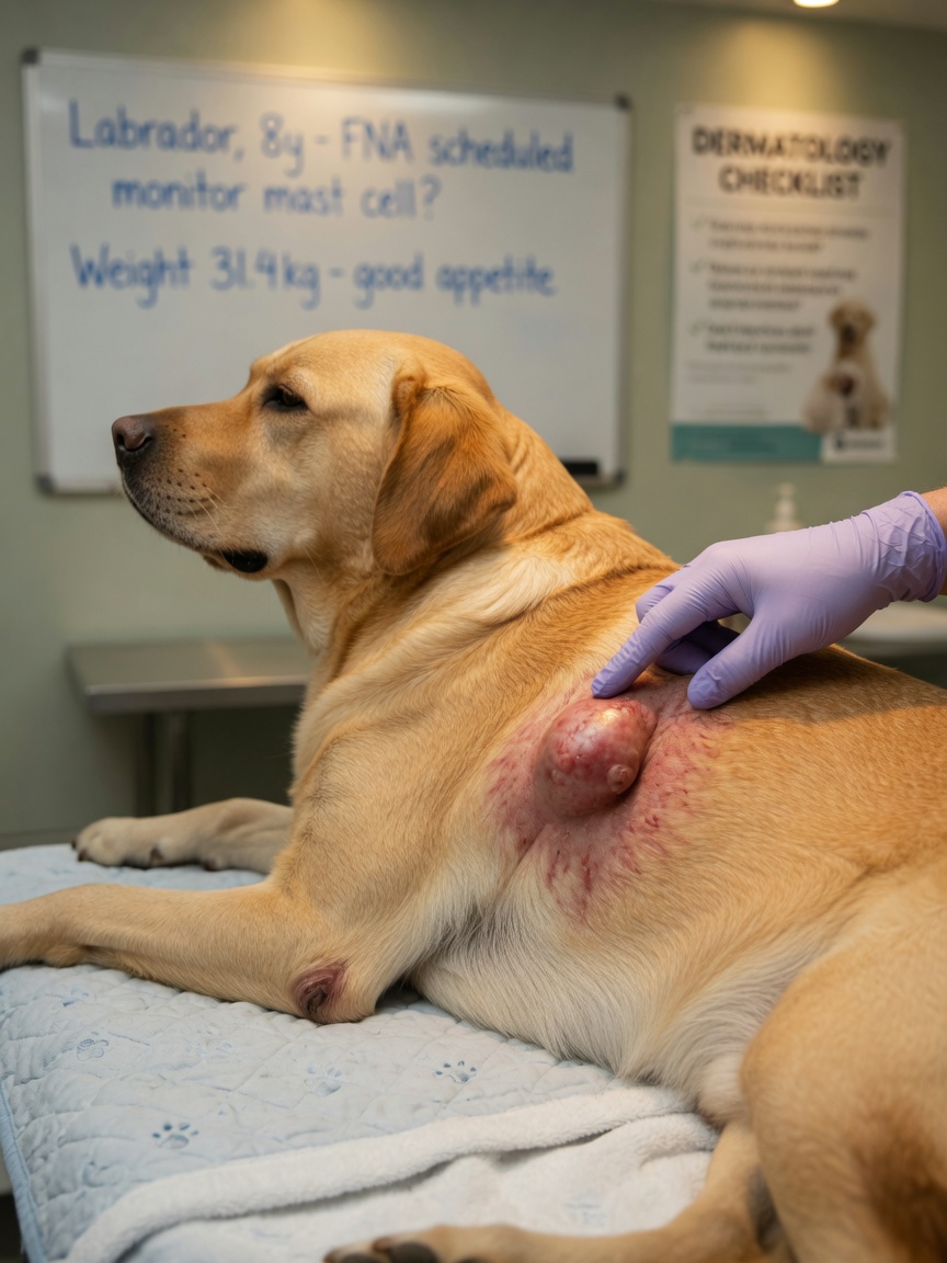
Mast cell tumours are notoriously variable in appearance, earning them the nickname the "great imitator." They can look like anything from a smooth raised button to an ulcerated, angry lesion. Many MCTs appear as a firm, raised skin lump that may have been present for months with little change, before suddenly altering in size.
A hallmark of mast cell tumours is their ability to change size. Manipulation of the tumour can trigger mast cell degranulation, causing the mass to swell, redden, and become surrounded by a flare of skin irritation — this is known as Darier's sign. A lump that becomes bigger and more inflamed when touched or stroked, and then subsides, is highly suspicious for an MCT and warrants prompt veterinary assessment. This fluctuation in size often leads owners to believe the lump has resolved, when in fact it has simply temporarily degranulated.
Grading: What It Means for Prognosis
The behaviour of mast cell tumours varies enormously, and grading systems help predict how aggressive a tumour is likely to be. Two main grading systems are used in veterinary oncology:
Patnaik System (Three Grades)
- Grade 1: Well-differentiated, confined to the superficial dermis. Excellent prognosis with complete surgical removal.
- Grade 2: Intermediately differentiated. Behaviour is variable and this grade can be difficult to predict — some behave like grade 1, others more aggressively.
- Grade 3: Poorly differentiated, deeply invasive. High metastatic potential and poor prognosis.
Kiupel System (Two Grades)
The newer Kiupel two-tier system classifies tumours as simply low grade or high grade based on specific histological criteria, and has been shown to be more reproducible and better at predicting outcome. High-grade tumours by the Kiupel system carry a median survival time measured in months without treatment, while low-grade tumours behave more benignly.
Your vet or a veterinary pathologist will advise on which grading system has been used and what the findings mean for your dog's individual case.
Surgery: The Primary Treatment
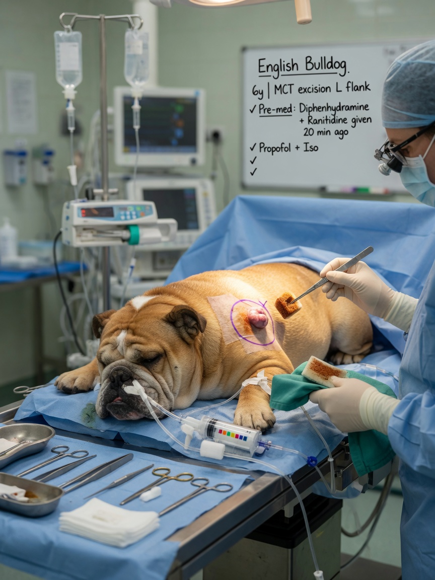
Surgical removal with wide margins remains the gold-standard treatment for mast cell tumours where this is achievable. The goal is to remove the tumour with a surrounding margin of normal tissue — typically at least two centimetres laterally and one fascial plane deep — to maximise the chance of complete excision. Incomplete excision significantly increases the risk of local recurrence.
Before surgery, veterinary surgeons commonly administer antihistamines and sometimes proton pump inhibitors. This pre-operative preparation is important because surgical manipulation of the tumour can trigger widespread degranulation, releasing large quantities of histamine into the bloodstream. This can cause acute hypotension, vomiting, and — in severe cases — anaphylaxis. Pre-medicating the dog reduces this risk substantially.
Stelfonta for Non-Resectable Tumours
For mast cell tumours that cannot be removed surgically — due to location, size, or anaesthetic risk — Stelfonta (tigilanol tiglate) is a licensed intratumoural injection that causes localised tumour destruction. It is approved in the UK for treatment of non-metastatic cutaneous mast cell tumours. The drug is injected directly into the tumour and triggers a process of localised cell death and immune activation, leading to tumour resolution in many cases. It is not suitable for all tumour locations and requires careful case selection by a veterinary oncologist.
Palladia for Metastatic Disease
Palladia (toceranib phosphate) is a tyrosine kinase inhibitor licensed for use in dogs with recurrent or metastatic mast cell tumours. It targets several molecular pathways involved in mast cell tumour growth, including c-KIT mutations which are found in a proportion of MCTs and are associated with more aggressive disease. Palladia is an oral medication given on an alternating-day schedule and requires careful monitoring for side effects including gastrointestinal upset, bone marrow suppression, and protein-losing nephropathy. It is typically administered under the supervision of a veterinary oncologist.
When Should You See a Vet?
Any new lump on your dog should be assessed by a vet. Lumps that change size, become red and itchy around their edges, ulcerate, or have been present for more than four weeks without investigation deserve prompt attention. A fine needle aspirate — a quick, minimally invasive in-clinic procedure — can often give an initial indication of whether mast cells are present, though histopathology after surgical removal is needed for definitive grading.
Early diagnosis and treatment significantly improves outcomes, particularly for higher-grade tumours where metastatic spread to lymph nodes and internal organs worsens the prognosis considerably.
Written by Sarah Bennett, pet health writer at ForPetsHealthcare.