
What Is a Lipoma?
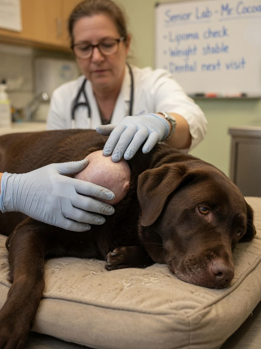
A lipoma is a benign tumour made up of mature fat cells (adipocytes). They sit within or just beneath the skin and are the most frequently encountered soft tissue tumour in dogs. Unlike many other lumps that develop on or under the skin, the vast majority of lipomas are entirely non-harmful — they do not spread to other parts of the body, they do not infiltrate surrounding tissue in their most common form, and they rarely cause the dog any discomfort unless they grow large enough to interfere with movement or become irritated by the dog's own grooming or lying habits.
Most lipomas feel soft and doughy to the touch, move freely under the skin when pressed, and have well-defined edges. They grow slowly, sometimes over the course of years, and owners often first notice them as a small pea-sized lump that gradually becomes more prominent.
Which Dogs Are Most Commonly Affected?
Lipomas become significantly more common as dogs age. They are rarely seen in dogs under five years old and are most frequently diagnosed in middle-aged to elderly dogs. Overweight dogs appear to develop lipomas more readily, although lean dogs can develop them too and weight alone is not a reliable predictor.
In terms of breed predisposition, Labrador Retrievers are particularly prone to developing multiple lipomas, sometimes accumulating a considerable number over the course of their lifetime. Other breeds commonly affected include:
- Cocker Spaniels
- Dachshunds
- Weimaraners
- Dobermann Pinschers
- Miniature Schnauzers
Female dogs may be slightly more predisposed than males, though lipomas occur in dogs of both sexes.
Infiltrative Lipomas: A More Challenging Variant
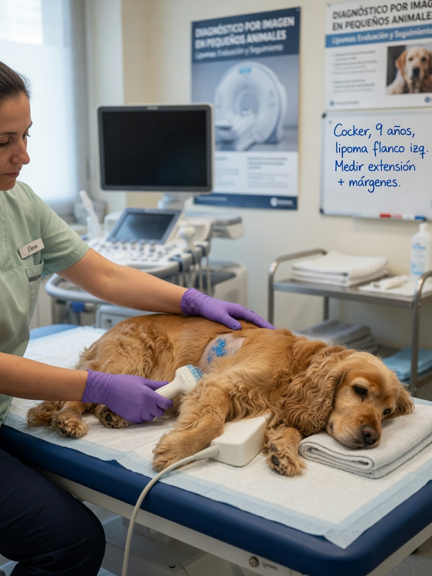
While most lipomas are straightforward subcutaneous fatty lumps, a variant known as the infiltrative lipoma behaves quite differently. Rather than sitting as a discrete, encapsulated mass beneath the skin, infiltrative lipomas grow into surrounding muscle tissue, fascial planes, and occasionally nerves. They do not metastasise — they are still locally benign in the sense that they do not spread via the bloodstream or lymphatics — but their tendency to invade between muscle fibres makes complete surgical removal extremely difficult.
Infiltrative lipomas are more likely to recur after surgical excision, sometimes multiple times, as small remnants of fatty tissue left behind during surgery can regrow. They are also more likely to cause functional problems, such as restricting a limb's range of motion or causing gait abnormalities, depending on their location. Imaging — typically MRI — is often needed before surgery to map the extent of an infiltrative lipoma and plan excision as thoroughly as possible.
Could a Lipoma Be Malignant?
Liposarcoma — a malignant tumour of fat cells — is rare in dogs, but it does occur. Liposarcomas are biologically distinct from lipomas; they are not thought to arise from existing benign lipomas but rather develop de novo. Nevertheless, owners and vets cannot distinguish a liposarcoma from a lipoma based on external feel alone. Liposarcomas tend to be firmer, less mobile, and more rapidly growing than typical lipomas, and they may be more deeply seated, but none of these features are definitive.
This is precisely why fine needle aspiration or histopathology is important for any lump that raises concern, rather than simply assuming a soft fatty lump is benign without investigation.
Fine Needle Aspirate: The First Diagnostic Step
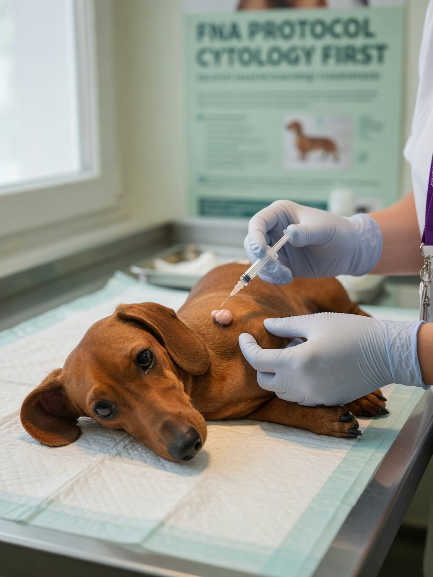
A fine needle aspirate (FNA) is a quick, inexpensive, and minimally invasive procedure that most vets can perform in a standard consultation. A thin needle is inserted into the lump and a small number of cells are drawn out and examined under a microscope. For lipomas, the aspirate typically shows large, clear fat droplets and mature adipocytes with no features of malignancy — a result that can reassure both owner and vet that a simple lipoma is present.
FNA is generally accurate for straightforward lipomas, though it is worth noting that some lumps yield poor or unrepresentative samples, and any lump with atypical features may warrant more formal surgical removal and histopathology for a definitive diagnosis. An inconclusive FNA result should prompt further investigation rather than watchful waiting if the lump is growing or changing.
When Should a Lipoma Be Removed?
The decision to remove a lipoma is not always straightforward and should be made on a case-by-case basis with your vet. Surgery is generally recommended in the following circumstances:
- Rapid growth — a lump that is noticeably enlarging over weeks to months warrants investigation and often removal
- Restricted movement — lipomas over a joint or axilla (armpit) that limit how the dog walks or sits
- Discomfort — if the lipoma is in a location where it is repeatedly compressed when the dog lies down or is knocked during normal activity
- Ulceration or skin breakdown — pressure from a large lipoma can cause overlying skin to break down
- Inconclusive FNA — if cytology cannot confirm benign nature
- Owner or vet preference when lump is small — smaller lipomas are easier to excise completely than large ones, and some vets recommend removal of confirmed lipomas while they are still manageable in size
Conversely, if a lipoma has been confirmed by FNA, is growing very slowly, and is not causing any functional problems, monitoring with periodic recheck appointments is a perfectly reasonable approach — particularly in elderly dogs for whom the anaesthetic risk of elective surgery needs to be weighed carefully.
What to Monitor Between Vet Visits
If you and your vet have agreed to monitor a confirmed lipoma rather than remove it immediately, keep a record of its size — a photo with a ruler beside it taken every few weeks is helpful — and contact your vet promptly if you notice any of the following:
- Sudden increase in size
- The lump becoming firmer in texture
- Loss of movement under the skin (becoming fixed to underlying tissue)
- The overlying skin becoming reddened or ulcerated
- Your dog showing any signs of discomfort when the area is touched
Lipomas in dogs are rarely a cause for serious alarm, but they should never be ignored entirely. Regular veterinary assessment of any lump, however innocent it appears, is always the safest approach.
Written by Sarah Bennett, pet health writer at ForPetsHealthcare.